|
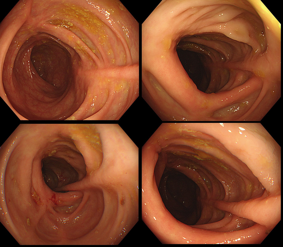
This is the colon of a 55 year old woman who has been treated for diarrhoea.
WHAT IS THE DIAGNOSIS?
■ Crohn's disease
Possible of course but why a 'middle aged lady'?
■ Ulcerative colitis
Zero points!
■ Microscopic colitis
Of course!
explanation
Actually, this patient had collagenous colitis and after treated with budesonide, symptoms settled. A colonoscopy 3 months later showed these linear scars throughout the colon.
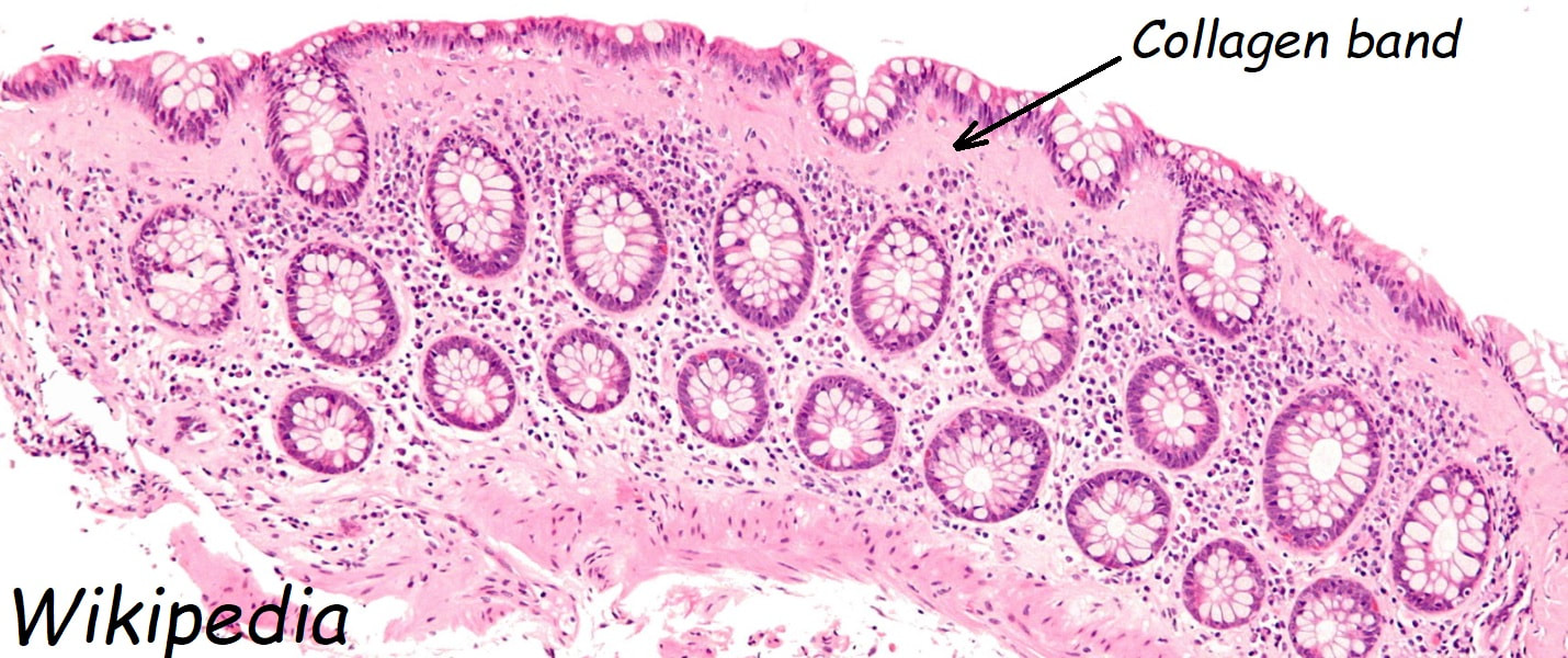
The first case of collagenous colitis was described in 1976 by Lindström and is characterised by chronic watery diarrhoea. Underlying hyperthyroidism, coeliac disease, bile salt diarrhoea and inflammatory bowel disease should be excluded. At colonoscopy, the mucosa is often unremarkable but there may also be mild, patchy erythema or linear cracks. Histology reveals thickening of the subepithelial collagen layer from the normal 3-6 μm to more than 10 μm as well as lymphocytic infiltration of the epithelium and the lamina propria (see image below). Rectal biopsies are not sufficient to make the diagnosis as normally the collagen layer is particularly thin here. Samples from the rectum and sigmoid confirms the diagnosis is more than 90% of cases. 9mg of Budesonide is effective but there is a high risk of relapse on stopping the drug [Bonderup OK. Gut 2003;52:248-51] and azathioprine is usually the second line agent. |
Categories
All
|


