Friends of Endoscopy
  • Home
  • Quiz Cases
  • Endoscopy Long Cases
  • Short Teaching Clips
  • Blackboard teaching
  • Podcasts
  • Core Reading
    • Basic concepts core reading
    • Gastroscopy core reading
    • Colonoscopy core reading
    • QA core reading
  • About
    • Our sponsors
  • Home
  • Quiz Cases
  • Endoscopy Long Cases
  • Short Teaching Clips
  • Blackboard teaching
  • Podcasts
  • Core Reading
    • Basic concepts core reading
    • Gastroscopy core reading
    • Colonoscopy core reading
    • QA core reading
  • About
    • Our sponsors
Search
Picture
​

Friends of Endoscopy is all about pattern recognition.  See it today and recognise it tomorrow!   Learn from a New Case on most weekdays !!! 
Become a Better Endoscopist ! 

A pancreatic mass

16/2/2021

 
Picture
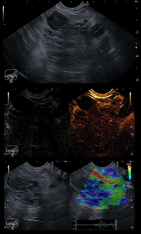
This is an contrast enhanced EUS of a 5cm pancreatic mass together with the doppler (arterial and venous phases) and elastography images
WHAT IS THE MOST LIKELY DIAGNOSIS?
■ Serous cystic neoplasm
Absolutely right !!!
■ Ductal adenocarcinoma
Nope !
■ Neuroendocrine tumor
No again!
■ Malignant intrapapillary mucinous neoplasm
No again!
explanation
This the typical appearance of a serous cystic neoplasm (formerly called serous cystic adenomas) situated in the tail of the pancreas. The natural history of these lesions is poorly known. 

The typical appearances includes:  
  • typical honeycomb appearance on gray-scale
  • hypervascularity on doppler
  • looks 'hard' on elastography due to the presence of multiple fluid filled spaces
Picture
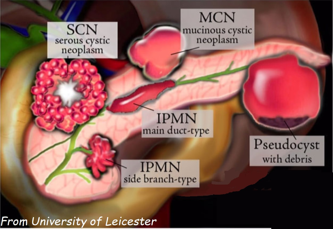
A multinational study of 2622 patients reported that these lesions are more common in women (1:4 ratio), avg age 58 yrs. In this cohort the size increased in 37% (growth rate: 4 mm/year), was stable in 57% and decreased in 6%. Only 3 serous cystadenocarcinomas were recorded and therefore a conservative approach was advocated. Most operations were carried out because of uncertainty over the diagnosis. Some would also consider offering surgery for patient with symptoms.  However, in the above series only 9% of patients presented pancreaticobiliary symptoms directly related to the lesion.  Because 10 patients died following surgery every effort must be made to make the diagnosis with the aid of CT, MRI and EUS and FNA. Low CEA levels with have an excellent specificity for diagnosis of a 'SCN' but can also be found in IPMNs and in neuroendocrine tumours. 

Prof Saftoiu and his team will be uploading further EUS cases in their new FB group 'Friends of Endoscopic Ultrasound' !  ​

    Categories

    All
    Barrett's
    Cancer
    Cancer Syndromes
    Colitis
    Colorectum
    Corrosive Ingestion
    Crypt Pattern
    Difficulty: Hard
    Difficulty: Moderate
    Difficulty: Very Hard
    Duodenum
    Eosinophilic Oesophagitis
    EUS
    Foveolar Metaplasia
    Gastric
    Gastroscopy
    GI Bleeding
    Histology
    HPB
    Ileum
    Immunosuppression
    Infection
    Local Recurrence Of Barrett's Ca.
    Lymphoma
    Mixed Polyp
    Mucosal Prolapse Syndrome
    NET
    Non-lifting Sign
    Oesophagus
    Opinion Piece!
    Pharynx
    Polyp
    Polypectomy
    The Basics
    TSA

  • Home
  • Quiz Cases
  • Endoscopy Long Cases
  • Short Teaching Clips
  • Blackboard teaching
  • Podcasts
  • Core Reading
    • Basic concepts core reading
    • Gastroscopy core reading
    • Colonoscopy core reading
    • QA core reading
  • About
    • Our sponsors