|
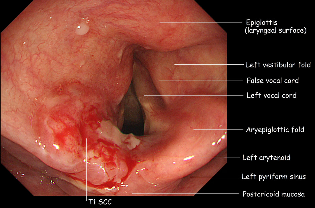
This lesion was found in a 65 yr old man complaining of odynophagia. WHAT IS THE LIKELY DIAGNOSIS?
■ Lympho-epithelial cyst
No, those are round, yellow submucosal swellings
■ Vocal cord papilloma
This is on the Cuneiform cartilage NOT the vocal cord!
■ Squamous neoplasia
There is a firm plaque of solid tissue and you can safely go one step further and guess that it's actually SCC
explanation
There is a firm, disc-like indurated area on the right aryepiglottic fold. This turned out to be a T1 SCC which was ultimately treated by radiotherapy.
Of course, we should not ignore this area on intubation! Take a moment before irritating the larynx with the tip of your endoscope to look at the larynx! Is everything symmetrical? Your most common finding will probably be a yellow, submucosal 'retention cyst, thought to be the result of retained mucus within a dilated mucus gland. However, particularly in smokers, it's vital to include this part of the endoscopy in your examination. Sadly, no endoscopy reporting system accommodate for reporting findings in this area. This makes it particularly pertinent that you learn what the landmarks are called !!! See below for an update and don't get confused between left and right ☺ ! |
Categories
All
|

