|
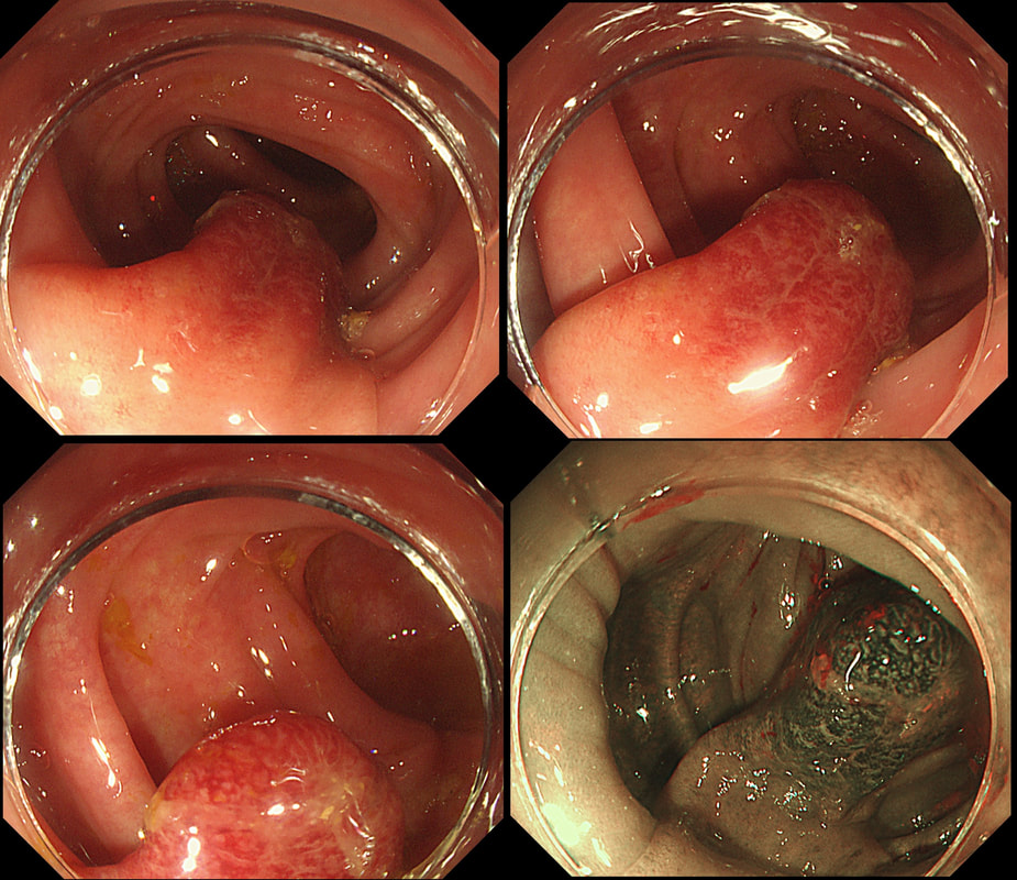
This polyp was found in the sigmoid colon. WHAT IS THE LIKELY AETIOLOGY OF THE POLYP? a) hyperplastic polyp b) adenomatous polyp c) malignant polyp Explanation
The sigmoid colon is the most 'powerful' part of the colon developing the force needed to go to the toilet. Presumably this is the reason why diverticular disease first develop in the sigmoid. The force can also create pseudo-polyps from patches of inflammation which I presume gets tugged along with each peristaltic wave. Even adenomatous polyps which develop here usually become traumatised. The end result is that this is the most difficult part of the colon to assess polyps. This particular lesion had been unchanged for several years but it spooked an endoscopist sufficiently to refer it for an EMR. Samples had indicated that it was a hyperplastic polyp only (correct answer was A). Fifteen years ago, the idea of subjecting the patient to an endoscopic resection for a lesion not recognised to be linked with cancer could have been criticised (if there was a complication). However, nowadays it is recognised that SSL's (hyperplastic polyps with L or T-shaped crypts etc) are 'fair game'. The truth is that it's probably impossible to tell a 'simple' hyperplastic polyp apart from a 'sessile serrated lesion' and pathologists are increasingly called all these 'serrated lesions'. For this reason, I go by the size. If the 'serrated lesion' is 10mm or larger, I usually remove it. In this particular 'polyp', most is actually just a swollen fold. The serrated area is situated at the very tip of the fold. When I see this, I don't go overboard placing the snare a long way down the 'pseudo-stalk'. If you did, you will find that it's taking a long time to cut through all that healthy sigmoid mucosal fold and you run the risk of a late perforation. Instead, I just caught the tip of the fold and ask my assistant to close the snare as quickly as possible whilst it was cut on a blend current (yellow pedal). Of course, you don't need to worry about large vessels in serrated polyps. |
Categories
All
|

