|
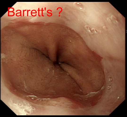
An endoscopic diagnosis of Barrett's has been made and this patient returns for further surveillance biopsies. Three years ago, no specialised columnar metaplasia was found.
DOES THIS PATIENT HAVE BARRETT'S?
■ No, I don't think so
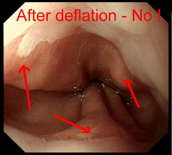
CORRECT, although it's easier to see when deflating that hiatus hernia!
■ Yes, probably!
INCORRECT, but deflate to make sure!
explanation
It can be difficult to tell if patients have Barrett's and I rather suspect that Barrett's is overdiagnosed at endoscopy. This is because you need to deflate the stomach to see the top end of those gastric folds. Below is the appearances after deflation of the stomach with the gastric folds reaching all the way up to the squamo-columnar junction. Remember, by definition, it's the top end of the gastric folds which marks the junction between the stomach and the oesophagus.
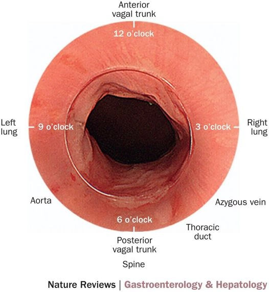
In most patients this should be where the squamo-columnar junction sits but of course, this moves proximally in Barrett's. How about taking biopsies to leave it to the pathologists to make the diagnosis? Few things upsets pathologists more than receiving some samples with the question: "- is this Barrett's?". The reason is that Barrett's is an ENDOSCOPIC diagnosis. Histologically, the normal cardia mucosa looks IDENTICAL to Barrett's and the pathologists can't tell the difference. OK! It's up to you to make that diagnosis and I've uploaded a second image below to remind you of the landmarks you should be aware of! |
Categories
All
|


