Friends of Endoscopy
  • Home
  • Quiz Cases
  • Endoscopy Long Cases
  • Short Teaching Clips
  • Blackboard teaching
  • Podcasts
  • Core Reading
    • Basic concepts core reading
    • Gastroscopy core reading
    • Colonoscopy core reading
    • QA core reading
  • About
    • Our sponsors
  • Home
  • Quiz Cases
  • Endoscopy Long Cases
  • Short Teaching Clips
  • Blackboard teaching
  • Podcasts
  • Core Reading
    • Basic concepts core reading
    • Gastroscopy core reading
    • Colonoscopy core reading
    • QA core reading
  • About
    • Our sponsors
Search
Picture
​

Friends of Endoscopy is all about pattern recognition.  See it today and recognise it tomorrow!   Learn from a New Case on most weekdays !!! 
Become a Better Endoscopist ! 

Five duodenal strictures

22/3/2021

 
Picture
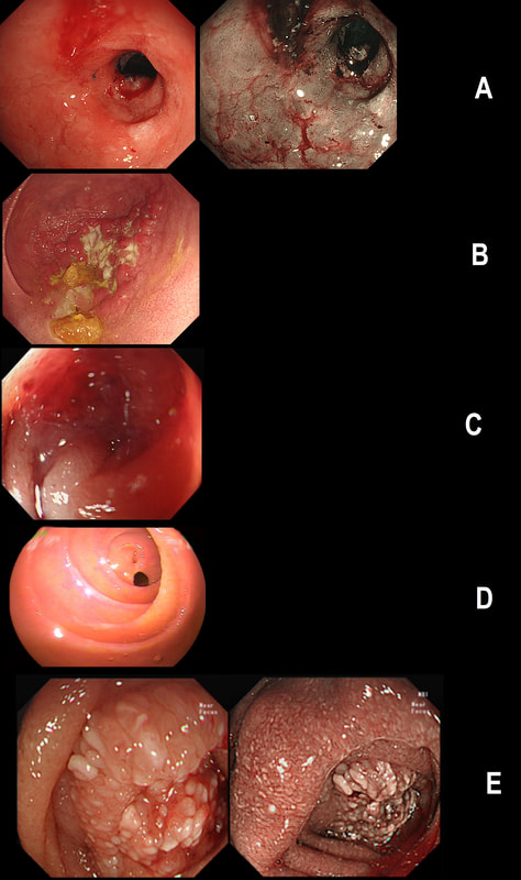
Just another piece of fun ☺!  Here are 5 photographs of duodenal strictures, each of a different aetiology! 
CAN YOU MATCH THE STRICTURE WITH THE PHOTO?
■ NSAID induced stricture =
Photograph D
■ Peptic stricture =
Photograph A
■ Chronic pancreatitis =
Photograph E
■ Chrohn's disease =
Photograph B
■ Duodenal adenocarcinoma =
Photograph C
EXPLANATION
You'll know that NSAID induced strictures are classically 'membrane-like' as in Photograph D. 

The 'Peptic stricture' is shown in photograph A.  The healed duodenal ulcer had been at 9 O'clock in that stricture.  

Chronic pancreatitis causes external compression of D1 or D2 usually with swollen villi. Of course there are two photographs to choose between - Photo C or E.  Lets leave that one and see if the Crohn's stricture is easier to find.

Of course, photograph B shows ulceration and inflammation and must be the CD stricture. 

That leaves the malignant stricture which could be photograph C or E.  It's usually relatively easy to squeeze past the external compression caused by a swelling in the head of the pancreas. In contrast, malignant duodenal strictures are usually impassable. Which one looks the tightest?  Photo C or E?  Well, C is the malignant stricture which I had to dilate up to 10mm before I could take samples a little deeper into the stricture to confirm the diagnosis.  Photograph E was a case of severe pancreatitis which we scoped as he had dropped his Hb (never found out why or how). 

    Categories

    All
    Barrett's
    Cancer
    Cancer Syndromes
    Colitis
    Colorectum
    Corrosive Ingestion
    Crypt Pattern
    Difficulty: Hard
    Difficulty: Moderate
    Difficulty: Very Hard
    Duodenum
    Eosinophilic Oesophagitis
    EUS
    Foveolar Metaplasia
    Gastric
    Gastroscopy
    GI Bleeding
    Histology
    HPB
    Ileum
    Immunosuppression
    Infection
    Local Recurrence Of Barrett's Ca.
    Lymphoma
    Mixed Polyp
    Mucosal Prolapse Syndrome
    NET
    Non-lifting Sign
    Oesophagus
    Opinion Piece!
    Pharynx
    Polyp
    Polypectomy
    The Basics
    TSA

  • Home
  • Quiz Cases
  • Endoscopy Long Cases
  • Short Teaching Clips
  • Blackboard teaching
  • Podcasts
  • Core Reading
    • Basic concepts core reading
    • Gastroscopy core reading
    • Colonoscopy core reading
    • QA core reading
  • About
    • Our sponsors