|
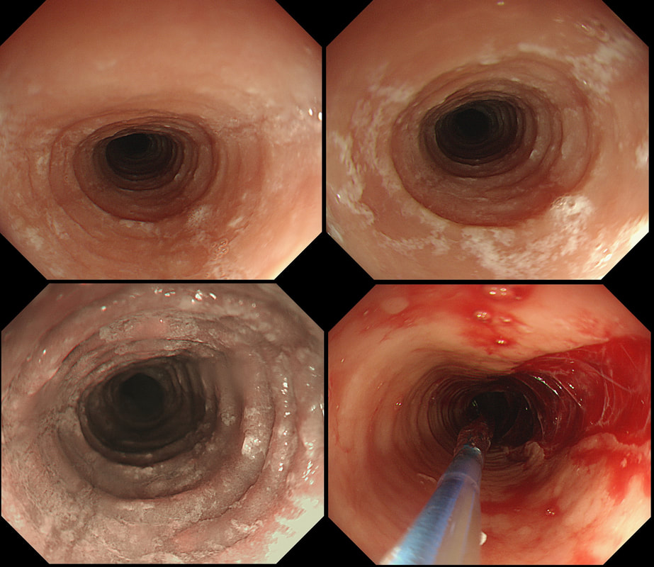
This patient had not responded to medication and was referred for a dilatation of this mid-oesophageal stricture.
WHAT IS AETIOLOGY OF THE STRICTURE?
■ Peptic
Nope, this doesn't look peptic
■ Candida
Hm, would not cause a stricture!
■ Pemphigus
Usually causes high oesophageal strictures
■ EoE
CORRECT!
■ Radiotherapy
Doesn't look like it!
explanation
This is a 'barndoor case'! A full house of eosinophilic micro-abscesses (those white spots which look like candida), longitudinal furrows and 'trachealisation', a rather soft sign of eosinophilic oesophagitis
It can by much more of a subtle diagnosis and for this reason you should always take oesophageal samples (1-2 samples at 2-3 different levels) in patients with odynophagia or dysphagia. Unfortunately, this patient did not respond to either a PPI, or oral budesonide but did feel better after the dilatation. By the way, this is what the mucosa normally looks like after dilating EoE. There is lots of fibrosis in the 'lamina propria' and I guess that's why it looks like this after a normal dilatation. |
Categories
All
|

