Friends of Endoscopy
  • Home
  • Quiz Cases
  • Endoscopy Long Cases
  • Short Teaching Clips
  • Blackboard teaching
  • Podcasts
  • Core Reading
    • Basic concepts core reading
    • Gastroscopy core reading
    • Colonoscopy core reading
    • QA core reading
  • About
    • Our sponsors
  • Home
  • Quiz Cases
  • Endoscopy Long Cases
  • Short Teaching Clips
  • Blackboard teaching
  • Podcasts
  • Core Reading
    • Basic concepts core reading
    • Gastroscopy core reading
    • Colonoscopy core reading
    • QA core reading
  • About
    • Our sponsors
Search
Picture
​

Friends of Endoscopy is all about pattern recognition.  See it today and recognise it tomorrow!   Learn from a New Case on most weekdays !!! 
Become a Better Endoscopist ! 

Two slides - one unifying diagnosis

1/3/2021

 
Picture
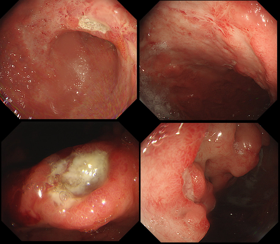
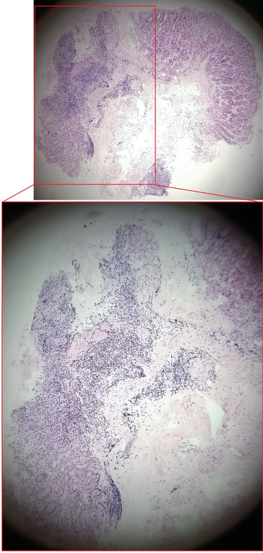
This ulcer was found on the lesser curve of a middle aged man with dyspepsia.  Biopsies are taken and stained with H&E (below)
Picture
WHAT IS THE AETIOLOGY OF THAT GU? 
■ Benign GU
Don't you think that the mucosa look odd near to the ulcer?
■ Lymphoma
Yes! That odd atrophic mucosa next to the ulcer look weird but not "carcinoma weird" !
■ Adenocarcinoma - intestinal type
With a 'standard cancer', the surrounding mucosa should be thickened and red not atrophic looking
■ Adenocarcinoma - intestinal type
This was actually my own diagnosis but histology shows lots of small round lymphocytes not ballooned up signet ring cells.
EXPLANATION
There are only two possibilities here.   Of course the ulcer itself doesn't tell you much. The mucosa surrounding the ulcer tells you more!   With a benign ulcer, you would expect a thin rim of 'reactive' mucosa (looks red and a little villous).  In the case of an intestinal type of adenocarcinoma a broad band of surrounding mucosa is red, indurated and thickened.   In gastric lymphomas, the surface area of abnormal mucosa is usually far greater than the surface area of the ulceration.  There are usually areas of atrophic and inflamed mucosa.   Mucosa infiltrated by a diffuse type adenocarcinoma could look like this BUT it's unusual for it to ulcerate!     

This patient has a MALT lymphoma !  Of course, I took samples for Helicobacters.  Of course not every MALT lymphoma is linked with HP infection (about 2/3 cases are though).  

In this case our haematologists were optimistic of a good response to Hp eradication because all the 3 good prognostic criteria were met:
  1. Only involvement of the mucosa (with no evidence of extension into the muscularis propria)
  2. No nodal disease on CT
  3. No 'unfavourable' mutations (such as translocations causing deregulation of MALT1 or Bcl-10 such as T(11;18), or overexpression of miR-142-5p and miR-155.

    Categories

    All
    Barrett's
    Cancer
    Cancer Syndromes
    Colitis
    Colorectum
    Corrosive Ingestion
    Crypt Pattern
    Difficulty: Hard
    Difficulty: Moderate
    Difficulty: Very Hard
    Duodenum
    Eosinophilic Oesophagitis
    EUS
    Foveolar Metaplasia
    Gastric
    Gastroscopy
    GI Bleeding
    Histology
    HPB
    Ileum
    Immunosuppression
    Infection
    Local Recurrence Of Barrett's Ca.
    Lymphoma
    Mixed Polyp
    Mucosal Prolapse Syndrome
    NET
    Non-lifting Sign
    Oesophagus
    Opinion Piece!
    Pharynx
    Polyp
    Polypectomy
    The Basics
    TSA

  • Home
  • Quiz Cases
  • Endoscopy Long Cases
  • Short Teaching Clips
  • Blackboard teaching
  • Podcasts
  • Core Reading
    • Basic concepts core reading
    • Gastroscopy core reading
    • Colonoscopy core reading
    • QA core reading
  • About
    • Our sponsors