Friends of Endoscopy
  • Home
  • Quiz Cases
  • Endoscopy Long Cases
  • Short Teaching Clips
  • Blackboard teaching
  • Podcasts
  • Core Reading
    • Basic concepts core reading
    • Gastroscopy core reading
    • Colonoscopy core reading
    • QA core reading
  • About
    • Our sponsors
  • Home
  • Quiz Cases
  • Endoscopy Long Cases
  • Short Teaching Clips
  • Blackboard teaching
  • Podcasts
  • Core Reading
    • Basic concepts core reading
    • Gastroscopy core reading
    • Colonoscopy core reading
    • QA core reading
  • About
    • Our sponsors
Search
Picture
​

Friends of Endoscopy is all about pattern recognition.  See it today and recognise it tomorrow!   Learn from a New Case on most weekdays !!! 
Become a Better Endoscopist ! 

What shape is the lesion?

26/5/2021

Comments

 
Picture
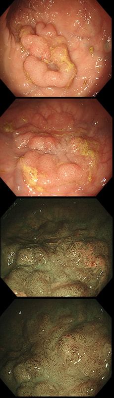
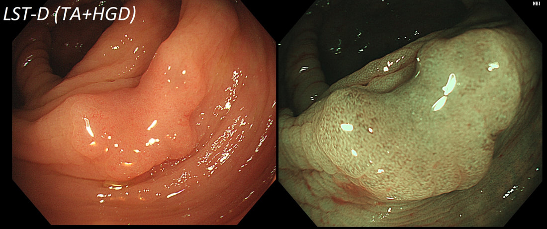
This lesion was found in the ascending colon.
HOW WOULD YOU DESCRIBE IT?
■ Flat elevated lesion (IIa)
Only lesions 10mm or smaller are referred to as 'IIa lesions'
■ Flat elevated lesion with a central depression (IIa+IIc)
But that depression dissappear when lumen is inflated!
■ Laterally spreading tumour of the granular type (LST-G)
Absolutely !
■ Laterally spreading tumour of the non-granular type (LST-NG)
But the surface is 'cobbled' not smooth!
■ Laterally spreading tumour with a central depression (LST-D)
LST-D's are always TA's (and therefore have a smooth surface)
explanation
Lets just state something from the start! A flat elevated polyp is of course a IIa type of lesion BUT when larger than 10mm in diameter is no longer referred to as a IIa lesion.  Now, it's a 'Laterally Spreading Tumour'!  
There are 4 types as follows:
  • LST-G (granular type) – usually found in the rectum or caecum and are almost always TVA’s harbouring LGD only
  • LST-NG (non-granular) – can be found anywhere in the colon and are always TA’s and usually harbour HGD (which should be your guess if the crypt openings are very small) or even cancer
  • LST-M (mixed type) – these are usually LST-G’s which have developed a dominant nodule. Resect the nodule first because if there is cancer, it will be found at the site of the dominant nodule
  • LST-D (depressed type) – this is the least common of all the LST’s are almost always TA’s in which early cancer has developed. 
I must admit to working from the crypt pattern and ‘backwards’ when deciding on the LST subtype.  Of course this is because the LST-G are always TVA’s with a gyrate crypt pattern whilst all the others are TA’s with slit-like crypts or small round crypts, usually found in LST-D's which usually harbour at least HGD.  This is because as the degree of dysplasia increase, the crypts wither, become smaller and more angular.   

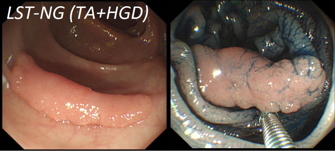
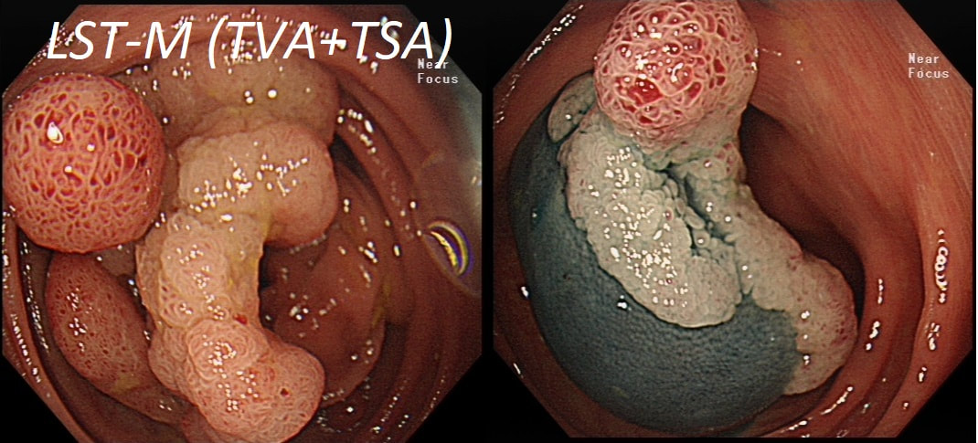
In this case the crypts are gyrate, (type IV crypts), making it a TVA. Laterally Spreading Tumours with a gyrate crypt pattern are all TVA's and almost never harbouring more than LGD.  OK but it looks depressed in the centre doesn't it?  No, it isn't depressed in the centre!  It's simply they way the large lesion has folded itself. This is something of a 'trick question' as I deliberately didn't inflate the colonic lumen fully in the first image.   When the lumen is inflated, you can see the true shape of the lesion; a LST-G ! 

A meta-analysis in 2013 by Voorham et al. [Voorham QJM. AM J Gastro 2013;108(7):1042-] concluded that pedunculated polyps were more likely to harbour KRAS mutation and APC mutations than flat lesions, and that flat lesions were more likely to harbour BRAF mutations.  Depressed lesions and LST-NG’s were particularly unlikely to contain KRAS.  

Below are some examples of LST's
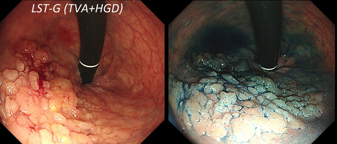
Picture
Picture
Picture
Picture
Comments

    Categories

    All
    Barrett's
    Cancer
    Cancer Syndromes
    Colitis
    Colorectum
    Corrosive Ingestion
    Crypt Pattern
    Difficulty: Hard
    Difficulty: Moderate
    Difficulty: Very Hard
    Duodenum
    Eosinophilic Oesophagitis
    EUS
    Foveolar Metaplasia
    Gastric
    Gastroscopy
    GI Bleeding
    Histology
    HPB
    Ileum
    Immunosuppression
    Infection
    Local Recurrence Of Barrett's Ca.
    Lymphoma
    Mixed Polyp
    Mucosal Prolapse Syndrome
    NET
    Non-lifting Sign
    Oesophagus
    Opinion Piece!
    Pharynx
    Polyp
    Polypectomy
    The Basics
    TSA

  • Home
  • Quiz Cases
  • Endoscopy Long Cases
  • Short Teaching Clips
  • Blackboard teaching
  • Podcasts
  • Core Reading
    • Basic concepts core reading
    • Gastroscopy core reading
    • Colonoscopy core reading
    • QA core reading
  • About
    • Our sponsors