Friends of Endoscopy
  • Home
  • Quiz Cases
  • Endoscopy Long Cases
  • Short Teaching Clips
  • Blackboard teaching
  • Podcasts
  • Core Reading
    • Basic concepts core reading
    • Gastroscopy core reading
    • Colonoscopy core reading
    • QA core reading
  • About
    • Our sponsors
  • Home
  • Quiz Cases
  • Endoscopy Long Cases
  • Short Teaching Clips
  • Blackboard teaching
  • Podcasts
  • Core Reading
    • Basic concepts core reading
    • Gastroscopy core reading
    • Colonoscopy core reading
    • QA core reading
  • About
    • Our sponsors
Search
Picture


Our short teaching videos are mini-learning opportunities for busy clinicians with a few minutes to spare!  The idea is to give you a quick reminder of issues, rather than a 30 minute in-depth review !  


Endoscopic resection of polyp invading diverticula – Tips/tricks/key points

5/9/2022

0 Comments

 
                                                                                                                                                                          Posted by Dr Pradeep Mundre
Picture
Picture
Picture
Picture
Picture
Picture
Picture
Picture
Picture
Picture
Picture
Picture


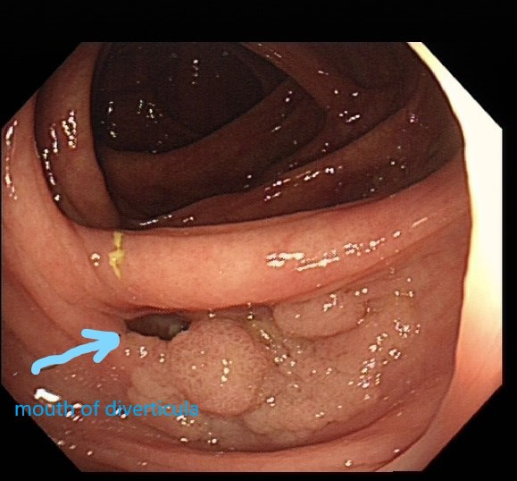
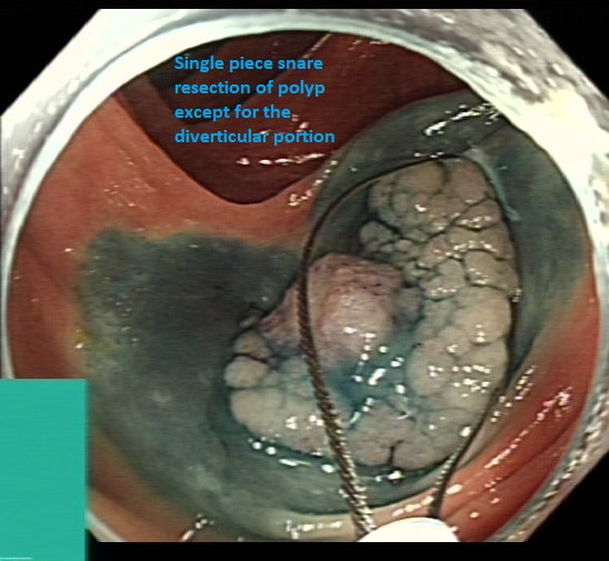
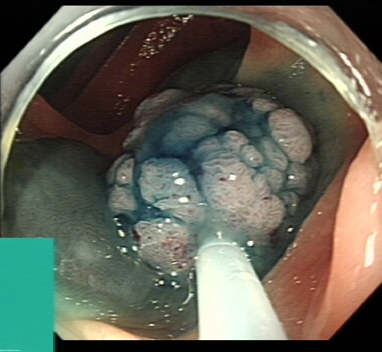
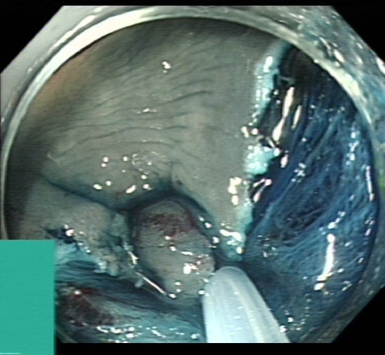
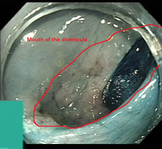
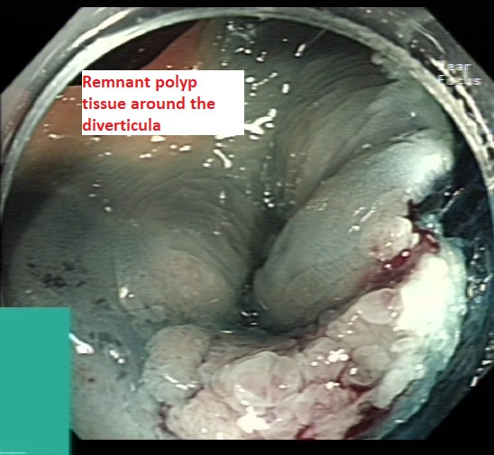
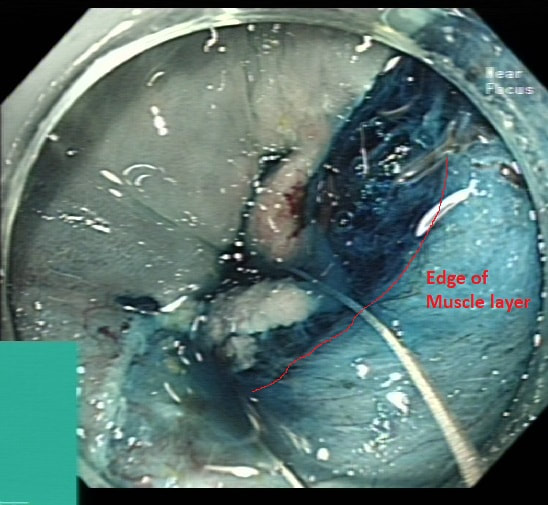
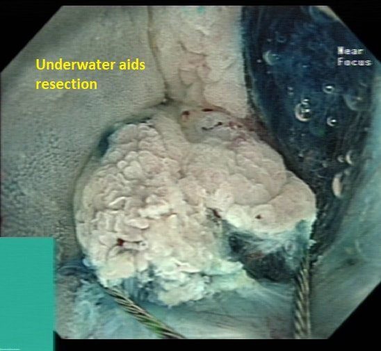
​Highlighting some key points, Tips/Tricks when considering endoscopic resection of polyps invading diverticula


1. More often the polyp just invades partially superficially into the diverticula, making it easy to tease it out during endoscopic resection
2. For most such polyps conventional EMR should be enough- Ensure that the lifting is in the right plane starting lift away from diverticula gradually moving towards.
3. There is a risk of perforation as no muscularis propria in diverticula
4. Clear cap always helps for any complex resection
5. There is often poor lifting of polyp around the diverticula
6. When resecting the polyp tissue within the diverticula, deflating the lumen and releasing the luminal tension helps
7. Stiff snare (I used medworks flat band snare here) may help get better hold of polyp
8. Underwater resection helps similarly
9. Despite macroscopically complete resection, risk of Recurrent/Residual polyp is high, so always consider site check @ 4-6 month and once again at 18 months
10. Other methods include EFTR, Band ligate and leave (post biopsy) approach or surgery
 
                                                                                                                                                                                           Dr Pradeep Mundre
0 Comments

Traction wire assisted ESD of a hepatic flexure LST NG  pseudodepressed

16/1/2022

0 Comments

 
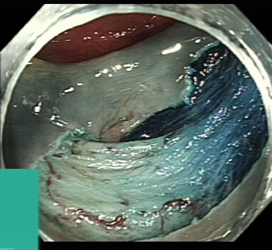
Short video of use of traction wire - Medtronic ProdiGI use in a ESD of a hepatic flexure LST NG type leaning towards pseudodepressed type , but can argue its flat type

Few points about LST NG Pseudodepressed type
  1. High risk of Sm invasive malignancy- approx 40 %
  2. Tend to have multifocal Sm invasion within the polyp
  3. Donot lift well with due to fibrosis
  4. Snare resection is difficult especially > 2 cms due to snare slippage
  5. One of the important indication for ESD - Enbloc resection
0 Comments

Piecemeal removal of 'difficult polyp' in ascending

19/4/2021

 
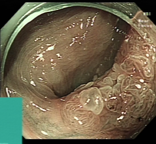
This is a 'difficult polyp because it's almost impossible to get any closer than this.  It's arising from a fold in the ascending colon, just downstream to the caecum.   Keep an eye on that scopeguide image and you see why it's so difficult. 

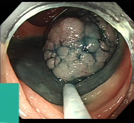
Of course this makes it difficult to tell how far the polyp extends into the caecal sac.  Fortunately, I was able to get glimpses of just behind the lesion which told me that it didn't extend far beyond the fold.   Had this lesion extended into the 'valley' behind that fold from which its arising, it would have been a 'show stopper' to me. 

Another reason to 'attack' is that the lesion does look benign with a reassuring gyrate crypt pattern of a TVA.  Naturally, any polyp of this size will have a 15% risk of actually containing an unexpected focus of cancer within.  Because our therapeutic waiting list is in mess in the aftermath of Covid, I do encourage everyone to sample the sessile lesion even though you are planning to refer it for resection. Such sampling on the surface of large sessile lesions doesn't induce any fibrosis and provides 'some' (very limited of course) reassurance that the lesion isn't malignant.  

     The main teaching points in the video are: 
  • It looks like a benign, sessile TVA and this was subsequently confirmed. 
  • The scope stability is an issue and it's difficult to get close to the polyp
  • Only a few can cope with >45min procedure and consider GA if you expect the resection to take longer in the right hemicolon 
  • On the left side, both you and your patient can take a break every 30-40 minutes . 
  • As you work and keep insufflating the ascending colon, the caecum will be pushed further and further away from you - be sparing with the insufflation!
  • A local recurrence will happen at that 'blind edge' of your resection
  • The muscle propria has taken a hammering and you must absolutely place clips both to reduce the risk of a late perforation but also to reduce the risk of bleeding

Syncronous/metachronous lesions

4/3/2021

 
Picture

Of course, its important to carefully examine the entire stomach especially in premalignant stomach for any synchronous lesions despite seeing something very evident.

The above video is follow up Endoscopy 3 months after the removal of the large sessile lesion in the antrum (photo).  

The polyp was removed by ESD and histology confirmed a Villous adenoma with low and high grade dysplasia as well as a small focus of adenocarcinoma – pT1aM2 (clear margins).

Wisely, it turned out, the patient was recalled for early 'site-check' and examination of the rest of the stomach.   This turned out to be worth while ! 

WHAT DO YOU THINK THE LESION IN THE VIDEO IS?

explanation
It is very easy to overlook synchronous lesions when there is a large 'eye-catching' lesion, drawing all your attention.  My learning point is that its important to look for synchronous and eventually metachronous lesions and not to get blinkered by the obvious pathology!   Patients do sometimes have dual pathology !  Yes, it happens !!! 

Actually, the second lesion was confirmed to be an Intramucosal adenocarcinoma - moderately differentiated pT1aM3, with no lymphovascular invasion and clear lateral and deep resection margins . This was a 'curative resection' as per BSG Guidelines for endoscopic resection as the risk of nodal metastasis in such leisons is probably <1%. 


Some bullet points on Synchronous tumours (Dysplasia or cancer)
  • Definition - Occurring at the same time diagnosed within 6 months (some use 1 year)
  • Incidence - Up to 30 % in those with gastric dysplasia
  • >50 % of gastric adenomatous polyps over 2 cm harbour adenocarcinoma

And some on the topic of Metachronous tumours 
  • Definition – tumours following in sequence – usually diagnosed more than 6 months apart (some use 1 year)
  • Incidence – about 3-15 % post ER over 5-7 years
  • Risk factors for metachronous lesions– persistent H pylori, age
  • Current advice is to resect all gastric Dysplastic lesions if appropriate due to risk of progression to malignancy
  • Most experts agree to offer long term surveillance for patients post Endoscopic resection and is usually undertaken annually
  • Most metachronous lesions can be successfully treated endoscopically

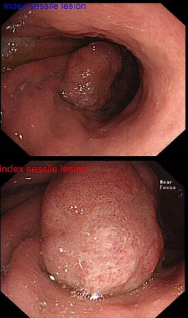
Reference
Banks M, Graham D, Jansen M, et al British Society of Gastroenterology guidelines on the diagnosis and management of patients at risk of gastric adenocarcinoma. Gut 2019;68:1545-1575.

Rectosigmoid LST-G: pEMR

7/2/2021

0 Comments

 
A 60 year old woman with type 2 diabetes and hypertension was found to be FIT+ve under the UK Bowel Cancer Screening Programme. The index colonoscopy, performed by a colleague 2 weeks ago, showed 4 polyps in the sigmoid / rectosigmoid colon. The three smaller polyps (6-15mm diameter) were removed (tubular adenoma, low grade dysplasia). The largest polyp, a 5cm LST-G  on a fold at the rectosigmoid junction was left behind for another day. The LST-G was not biopsied. The patient now attends for a further procedure to remove the LST-G (see video…).


The video highlights several tips:
  1. Using a gastroscope to improve access in a tricky area of the bowel.
  2. Using retroflexion to get better views of a polyp draped over a fold.
  3. Using good volumes of lift solution to get a decent cushion.
  4. Using gravity to your advantage by rotating the patient.
  5. Extending the defect contiguously and systematically.
  6. Using Coagrasper to control bleeding.
  7. Using snare-tip Softcoag to treat the edges of the defect.
0 Comments

Rembacken's Resection Rules

7/2/2021

 
'Rembacken's Resection Rules' is a 'tongue in cheek' list of General Principles which helps me in my endoscopic practise.   They have stood the test of time !!! 

Contralateral burn in a spastic sigmoid

4/2/2021

0 Comments


Sigmoid snare polypectomy in spastic, tight sigmoid. Teaching points include:
  • Use a Gastroscope
  • Removal in retrovertion can be impossible
  • Consider buscopan for spasm
  • Allow 40-60 minutes
  • Full colon bowel cleansing
  • Keep snare away from mucosa
  • Recognise collateral burns and apply clips

0 Comments

Snare polypectomy

3/2/2021

0 Comments


Small polyp on thick stalk for snare polypectomy at the recto-sigmoid junction cocked up! 
Teaching points include:
  • Don't rush it! 
  • Bring polyp down to 6 O'clock or resect in retrovertion
  • Assess snare position before cutting 
  • Immediate bleeding is easy to deal with
  • Assess for complete excision
  • Consider clips 
0 Comments

EMR of 20mm sigmoid polyp

2/2/2021

1 Comment


EMR of a 20mm sigmoid polyp.  Main teaching points includes:
  • Use a gastroscope
  • Removal in retrovertion is easier
  • Give full colonic prep
  • Keep the resection area clean and dry
  • Anticipate bleeding before it happens
  • Deal with bleeding straight away
  • Place the first clip on the most awkward spot
1 Comment

Lugol's iodine to map squamous dysplasia

2/2/2021

0 Comments


A short clip of Lugol's dye spray. Teaching points includes:
  • Squamous dysplasia is well seen by blue light technologies
  • First wash with lots of acetylcysteine
  • Consider topical lignocaine but don't mix it with Lugol's
  • Dilute the 5% Lugol's down to 1%
  • Flat squamous dysplasia can usually be resected whilst nodules usually can't be
0 Comments

    Categories

    All
    Bleeding
    Colorectum
    EMR
    Gastroscopy
    Oesophagus
    Rembacken's Rules
    Snare Polypectomy
    Squamous Mucosa

    Archives

    January 2022
    April 2021
    March 2021
    February 2021

  • Home
  • Quiz Cases
  • Endoscopy Long Cases
  • Short Teaching Clips
  • Blackboard teaching
  • Podcasts
  • Core Reading
    • Basic concepts core reading
    • Gastroscopy core reading
    • Colonoscopy core reading
    • QA core reading
  • About
    • Our sponsors