Friends of Endoscopy
  • Home
  • Quiz Cases
  • Endoscopy Long Cases
  • Short Teaching Clips
  • Blackboard teaching
  • Podcasts
  • Core Reading
    • Basic concepts core reading
    • Gastroscopy core reading
    • Colonoscopy core reading
    • QA core reading
  • About
    • Our sponsors
  • Home
  • Quiz Cases
  • Endoscopy Long Cases
  • Short Teaching Clips
  • Blackboard teaching
  • Podcasts
  • Core Reading
    • Basic concepts core reading
    • Gastroscopy core reading
    • Colonoscopy core reading
    • QA core reading
  • About
    • Our sponsors
Search
Picture


Our short teaching videos are mini-learning opportunities for busy clinicians with a few minutes to spare!  The idea is to give you a quick reminder of issues, rather than a 30 minute in-depth review !  


Syncronous/metachronous lesions

4/3/2021

 
Picture

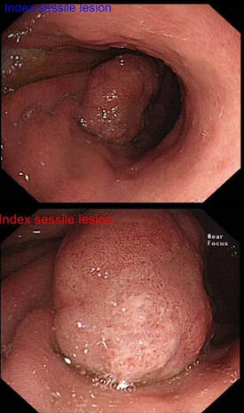
Of course, its important to carefully examine the entire stomach especially in premalignant stomach for any synchronous lesions despite seeing something very evident.

The above video is follow up Endoscopy 3 months after the removal of the large sessile lesion in the antrum (photo).  

The polyp was removed by ESD and histology confirmed a Villous adenoma with low and high grade dysplasia as well as a small focus of adenocarcinoma – pT1aM2 (clear margins).

Wisely, it turned out, the patient was recalled for early 'site-check' and examination of the rest of the stomach.   This turned out to be worth while ! 

WHAT DO YOU THINK THE LESION IN THE VIDEO IS?

explanation
It is very easy to overlook synchronous lesions when there is a large 'eye-catching' lesion, drawing all your attention.  My learning point is that its important to look for synchronous and eventually metachronous lesions and not to get blinkered by the obvious pathology!   Patients do sometimes have dual pathology !  Yes, it happens !!! 

Actually, the second lesion was confirmed to be an Intramucosal adenocarcinoma - moderately differentiated pT1aM3, with no lymphovascular invasion and clear lateral and deep resection margins . This was a 'curative resection' as per BSG Guidelines for endoscopic resection as the risk of nodal metastasis in such leisons is probably <1%. 


Some bullet points on Synchronous tumours (Dysplasia or cancer)
  • Definition - Occurring at the same time diagnosed within 6 months (some use 1 year)
  • Incidence - Up to 30 % in those with gastric dysplasia
  • >50 % of gastric adenomatous polyps over 2 cm harbour adenocarcinoma

And some on the topic of Metachronous tumours 
  • Definition – tumours following in sequence – usually diagnosed more than 6 months apart (some use 1 year)
  • Incidence – about 3-15 % post ER over 5-7 years
  • Risk factors for metachronous lesions– persistent H pylori, age
  • Current advice is to resect all gastric Dysplastic lesions if appropriate due to risk of progression to malignancy
  • Most experts agree to offer long term surveillance for patients post Endoscopic resection and is usually undertaken annually
  • Most metachronous lesions can be successfully treated endoscopically

Reference
Banks M, Graham D, Jansen M, et al British Society of Gastroenterology guidelines on the diagnosis and management of patients at risk of gastric adenocarcinoma. Gut 2019;68:1545-1575.

Comments are closed.

    Categories

    All
    Bleeding
    Colorectum
    EMR
    Gastroscopy
    Oesophagus
    Rembacken's Rules
    Snare Polypectomy
    Squamous Mucosa

    Archives

    January 2022
    April 2021
    March 2021
    February 2021

  • Home
  • Quiz Cases
  • Endoscopy Long Cases
  • Short Teaching Clips
  • Blackboard teaching
  • Podcasts
  • Core Reading
    • Basic concepts core reading
    • Gastroscopy core reading
    • Colonoscopy core reading
    • QA core reading
  • About
    • Our sponsors