|
|
Our podcasts give you an update on the latest Endoscopy related developments. A new episode is launched every few weeks. Listen on the Podcast app of your choice !
|
Transcript
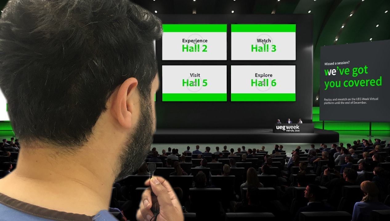
Once you had registered you were met by large entrance hall where you could click on: 1) World of Science 2) Industry exhibition 3) My UEG Community 4) TV study 5) Missed a Session – repository of past sessions 6) Industry Symposia 7) Press 8) Help desk 9) Link to the app World of Science gave you access to 6 ‘Halls’ · All presentations had been pre-recorded but they felt ‘live’ as the session was introduced and managed by chairmen just like a normal meeting. · During and after the presentations there was an opportunity to ask questions which were first vetted and then uploaded for all to see · There was a total of 626 presentations, delivered during 177 sessions by 502 speakers as well as >1400 E-posters. · The IT side held up very well and all presentations streamed with good sound. Sometimes it could be a little difficult to see the slides, even on a larger screen. Of course, all the chairmen were actually sitting at home and occasionally their own WiFi signal dropped making the feed jerky. However, this was far less of a problem than when taking part in a MS Teams meeting at work Screening 1) Early gastric cancer – 2) Marco Dinis-Ribeiro from Porto reminded us about the MAPS guidelines. He quoted several papers to claim that we can diagnose gastric intestinal metaplasia endoscopically without need for histological confirmation; § Pimentel-Nunes P. Endoscopy 2016;48(8);723-30, § Ang TL. J Gastroenterol Hepatol 2015; 27: 1473–1478, § Lage J. Scand J Gastroenterol 2016; 51: 501–506, Marcos P. Gut 2019;69;1762-8 3) Basically, it’s things like NBI or BLI which increases the endoscopic detection rates from quote poor 50% to close to 90%. Gastric atrophy on the other hand remains difficult to diagnose both endoscopically and histologically 4) Whether this holds true on busy, normal jobbing endoscopy lists, outside of specialist centres has not yet been proven. § reminding us of the MAPS II guidelines of 2019 by Marco Dinis-Ribeiro. · Questions included is it cost effectiveness of doing 3-yearly surveillance to patients with OLGIM 4 changes – uncertain · Do we need to always sample or can we make an endoscopic diagnosis of intestinal metaplasia? He claims that we can ! Mario quoted several papers in support of this: Pimentel-Nunes P. Endoscopy 2016;48(8):723-30, Castro R. Scand J Gastro 2019;54(11):1301-1305.) and Marcos P. Gut 2019 § You may have come across ‘hereditary gastric cancer’? You will suspect this if: · A pt develops diffuse gastric cancer before 40 · 2 family members with diffuse type gastric cancer. · history of diffuse lobular breast cancers · Pt has a cleft lip or palate § Of course the common syndromes are 1) hereditary diffuse type gastric cancer (pts have a mutation in the e-cadherin genes), Peutz-Jegherz syndrome and Juvenile polyposis syndrome (no strong link with FAP) § In pts with e-cadherin mutation targeted biopsies of any suspicious lesion as well as a minimum of 30 mapping random biopsies should be taken (the Cambridge endoscopy protocol). § Time-consuming, tedious process, which significantly prolongs the procedure and might reduce patient tolerance. In order to save time two specimens can be taken during a single passage of the forceps ("double-bite" technique). § II recall one such patient in Leeds § Now there was an abstract (P1138) on patients with ‘hereditary diffuse type of gastric cancer’ in which 51 confirmed carriers underwent surveillance. Both the endoscopist and the biopsies missed cancer in 23 pts. If you exclude the cases when the endoscopist saw nothing but the histopathologist spotted the cancer in one of the <70 random biopsies (!), the miss-rate of endoscopy was 73% !!! The authors concluded that either they needed to take more samples (1700 in fact) or surveillance simply doesn’t work in this group. No shit ! 5) Barrett’s surveillance § Reminded us to always retroflex and have a look at the GOJ from below § Brushings (Wide Area Transepithelial Sampling = WATS, Vennalaganti GIE 2018;87(2):348-55) samples larger areas but requires AI to detect markers for dysplasia or for risk and takes an extra 4.5 minutes (pretty much doubles the duration of the examination). § The recent Lancet paper by Rebecca Fitzgerald was mentioned; you may already be familiar with this paper which indicated that risk of future cancer could be estimated 8-10 years into the future by looking at number of cells with multiple sets of DNA · There was an update from Cambridge (A. Katz-Summercorn, OP016) who reminded us that there is no difference in the ‘gene copy number’ (the number of clonal mutations) between Barretts which progresses and does not progress to dysplasia and cancer. Instead it was the ‘genomic heterogeneity’ which predicted risk of progression. Patients who progressed had a) more translocations whereby parts of chromosomes had broken off and re-attached itself at the wrong site, b) a larger number of mutations, c) ‘driver gene alterations’ (p53 mutations etc ) · Unfortunately, the genomic heterogeneity’ changed in step with the dysplasia rather than before the dysplasia developed · Question arises of RFA can really be expected to ‘re-set’ the clock and if the same chromosomal translocations happens throughout our bodies as we age (from 30 translocations in the genome of an ‘indolent case of Barrett’s to ‘chromoplexy’ where more genes seem to be linking up with the wrong chromosome and finally ‘chromotrypsis’ where the genome is essentially ‘shattered’ and the patient has IMca. · Would it be expensive to do this ‘whole genome sequencing’ of DNA extracted from Barrett’s samples and give a single figure for the ‘genomic heterogeneity’. § We were reminded of the Dutch study published in Am J Gastro in 2010 by Curvers et.al. and subsequently by Duits (Gut 2014) that 9-13% of pts with Barrett’s LGD progress to HGD or IMca each year compared. But that unfortunately, only 1:7 to 1:4 pts originally diagnosed with LGD was on review confirmed as actually having LGD. · We are supposed to have all diagnoses of LGD confirmed by an ‘expert pathologist’. But how is this defined ??? · Dr N Frei at Amsterdam University Medical Center reported (OP017) on the 155 pts with Barrett’s LGD who took part in the SURF Trial (remember this is the study which reported progression in 20% (34/155). Using a pathological immunohistochemistry stain and software called ‘tissue cypher) · 8% of patients which ‘TissueCypher’ called ‘high risk’ progressed. · My suspicion is that TissueCypher got it right and the pathologists got it wrong !!! · This was supported by the finding that a panel of 3 pathologists who reviewed the diagnosis of LGD in these 155 pts only agreed in 50% of cases and 1/3 of patients were downgraded as not having any dysplasia at all !!! · I was going to phone the pathology department asking them to purchase that ‘TissueCypher’ when I heard that everything hinges on the CORRECT histological slides being submitted for ‘TissueCypher’ analysis! § The basic question of which patient with reflux should be offered an endoscopy to check for reflux – remains unanswered. Because about 15% of adults do have intermittent reflux and we are all overweight ! § Should all patients with Barrett’s be started on a PPI. Roos’s answer was YES as there is evidence that it may reduce the risk of progression to dysplasia. However, as far as I understand the risk of progression once you have already developed dysplasia is not affected by the use of PPI ? 6) Surveillance to prevent Pancreatic cancer in high risk groups? § Obviously not for everyone but perhaps in high risk groups with: a) new onset diabetes, b) pts found to have a pancreatic cyst or c) those with a family history or a known mutation such as CDKN2A (mutation carriers get familial melanoma, glioblastoma and pancreatic cancers), BRCA1 or 2, MLH1 or MSH2 or MSH6 and pts with Peutz-Jeghers syndrome etc. It turns out that there is no evidence that regularly surveilling these patients saves lives. 7) Colorectal cancer § Polish study reporting that the lowest uptake of screening was in the countries where colonoscopy was done under propofol. Actually, it was lowest in the countries were patients when straight for colonoscopy without a faecal test first. The study found that those >60 yrs, living <20 km from screening centre and first having been offered a FIT were more likely to accept screening. GI bleeding · Retrospective study of OVESCO clip vs angiography in Germany, · 1426 GI bleeding cases and only 128 pts were selected (highly selected) · Outcomes were similar in the over-the-scope clip group vs those who underwent angiography. The only thing which was significantly different was the in hospital mortality which was higher in the angiography group – understandably !!! · We were reminded (IP155) of another prospective study of OVESCO clip in UGI bleeding, called the ‘Sting trial’ (Schmidt A. Gastroenterology 2018;155(3):674-86), in which 5 centres in Germany, Switzerland and Hong Kong recruited a total of 63 patients over a 3 year period who had re-bled from a peptic ulcer. 1) Half were then treated with ‘standard therapy’ (and which wasn’t standard as patients had adrenaline+clip as ONLY 2 pts had adrenaline+heat) vs the over-the-scope clip. 2) 14/33 continued to bleed when their ulcer was re-treated with adrenaline+more clips vs only 2/33 in the OVESCO arm. 3) I have some concerns about the study: § In Leeds we published our experience on upper GI bleeding in 2016 and we had a total of 48 recurrent peptic ulcer bleeds over a 4 year period (End Int Open 2016;4:E282-6). You would have expected these 5 centres to generate more than only 66 pts over a 3 yr period § In fact, you would have expected there to be twice as many in the study patients § I think that the authors may have been concerned at the onset that some ulcers may not be ‘suitable’ for the OVESCO clip as the initial protocol excluded patients with “endoscopic failure to reach the bleeding source” § That not every patient was randomised was also suggested by the fact that “All endoscopic procedures in each center were done by 1 to 2 experienced endoscopists”. Is it really possible that 2 senior doctors were on-call for 24 hours, every other day for 3 years to be available when a patient suffered re-bleed ? § I am also concerned that the ‘standard’ treatment arm did not receive standard therapy § Finally, the study was not independent as the principal author received payment by OVESCO for consulting, lectures and research grants · An intermediate report on an ongoing prospective study (abstract P0041) from Korea of the use of a new haemostatic powder which they called ‘Nexpowder’ in patients with peptic ulcer bleeding. 11 patients (out of 70) re-bled after ‘standard dual therapy’ compared with 3 patients (out of 71) in the Nexpowder group. I was going to make an order for that stuff when it was pointed out that actually half of the 11 patients who re-bled after conventional therapy ONLY had adrenaline injected into their ulcer. It seems that the gold standard of ‘Dual Therapy’ for bleeding peptic ulcers is not commonly recognised in Korea. · Dr R. Inchingolo (IP069) reminded me of the Oakland score (Oakland K. Gut 2019;68:776-89) dividing patients with stable (=Shock index >1) with lower GI bleeding into high and low risk (includes age, sex, previous admission, blood on PR, HR – MOST important, systolic BP and Hb). Patients with PR bleeding and a shock index > 1 (HR/systolic BP) went directly to angiography. 1) Patients with a shock index and an Oakland score of 8 or less was discharged (eg. A 70 yr old woman with blood on PR but with a heart rate of 70 and no hypotension and normal Hb). 2) My concern with the BSG guidelines is that patients with a higher score should be offered a colonoscopy at the next available list. In my experience it’s usually impossible for these elderly and frail patients to take sufficient prep for this colonscopy to be worthwhile. § The BSG guidelines do admit that there ‘is no evidence for a benefit of colonoscopy’ in patients with lower GI bleeding. Of course this is hard to deny as even a meta-analysis of the 4 randomised trials conducted (Tsay C et.al. Clinical Gastroenterology & Hepatology 2019 - https://doi.org/10.1016/j.cgh.2019.11.061), reported that there was no benefit in mortality (RR, 0.93; 95% CI, 0.05–17.21), diagnostic yield (RR, 1.09; 95% CI, 0.99–1.21), endoscopic intervention (RR, 1.53; 95% CI, 0.67–3.48), or any primary hemostatic intervention (RR, 1.33; 95% CI, 0.92–1.92) with early colonoscopy (<24 hrs) even in severe cases of lower GI bleeding. Not surprising as about 95% of lower GI bleeding will stop spontaneously, 1/3 will rebleed once and ½ will rebleed a third time (Lim J. Tech Vasc Interv Radiol 2005) !!! · A very large study from India reported on the outcomes of 439 pts with moderate-severe radiation proctopathy. Success was declared in 403/439 (97%) after only a few APC sessions. Failure was linked with mucosal ulceration, >10cm long segment or >60% of circumference was involved. However, there were 6 severe adverse events which included rectal perforations and recto-vaginal fistula. There was a risk of strictures as well but this was not quantified. Adverse events were linked with mucosal ulceration … However, the patients treated with APC was those who had failed sucralfate. Sucralfate is still the mainstay of therapy, thank God. Therapeutics 1) There was a session on oesophageal strictures where a few interesting points were raised: § A patient presenting with a food bolus obstruction and have features of EoE. Would you carry out a dilatation at the index procedure? The answer was no because topical steroids will usually have an effect within 3-5 days § How long can you leave a patient with a food bolus obstruction or if a battery is stuck in the oesophagus or even if it’s just laying in the stomach? The ESGE guidelines say ≤6 hours § In some units the administer IV glucagon which supposedly have a 50% success rate. Not recommended by the ESGE § A new device was mentioned called the ‘excavator’ – a plastic over-the-scope grasper § Of course what do try is first to push the bolus into the stomach and if this fails, try grasping with either a basket or perhaps a snare § Manning wasn’t keen on the use of an overtube and preferred a large flexible hood fitted to the end of the scope. 2) Prof Inoue gave a presentation (IP017) of POET (Peroral Endoscopic Tumour Resection) whereby lesions attached to the muscle propria layer can be resected using a tunnelling technique. Looking at his footage, I was reassured that Prof Inoue could point out the vagus nerve, the recurrent laryngeal nerve and other important landmarks to me. Do I want to spend time dissecting nerves away from benign, submucosal lesions? 3) The OVESCO clip has been used for full thickness resections in the upper GI tract. In the stomach, R0 resection rates was 75%, complication rates was 30%. § When know that in the colon, full thickness resection is linked with a substantial perforation rate and an R0 rate of only about 70% and in lesions which are larger than 2cm it drops to 60%. Therefore its recommended that only lesions up to 2cm should be considered for this. § Of course that staging MRI would have to be done before that clip is placed and that the TME plane will be difficult to assess if the patient goes on to further surgery. 4) GERDx (Endoscopic Plicator) can be used to resect larger lesions <4cm in the stomach. R0 resection in 85% 5) An International Group was reached a non-controversial consensus on papillectomy (ampullectomies). Contraindications included extension into CBD of >10mm. I would have put the figure lower because EUS usually underestimate this 6) A French multi-centre study presented by Dr R Hallit (OP051) reported a 90% success rate treating early leaks with stents or pig-tailed stents. No mention of ‘vacuum therapy’ !? 7) Fortunately, in the same session there was a report (P. Stathopoulos OP052) of the immediate use of EVT in 9 cases of perforation (because the patient had swallowed something sharp or perforation complicating a dilatation). Immediately placing a suction catheter in the oesophageal lumen closed all 9 defects within 1-3 weeks. They replaced the sponge every 3 days but with the suction catheter placed within the oesophageal lumen, I don’t quite understand why this would be necessary?! 8) Another study in 24 pts with gastroparesis (21 being female and only 6 had diabetes?!) all treated with G-POEM (Gastric Peroral Endoscopic pyloroplasty) in Holland (Conchilo JM OP055) reported some improvement in 14 pts and no improvement in 10 pts. There was no way of predicting who would improve. The study group looked at the ‘pyloric distensibility index’ measured by ‘Endoflip’ or the ‘antro-duodenal manometry pattern’. § Perhaps not surprising because delayed gastric emptying could be due to non-relaxation of the pylorus, or poor contractility of the antrum, slow transfer of food by the duodenum or perhaps relaxation of the gastric fundus. § A sham study is required next to prove that G-POEM is better than placebo § Artificial intelligence · Fujifilm has CAD-EYE · Medtronics – GI Genius · Olympus – EndoBrain · Pentax – Discovery AI · There are others such as AI-Wision, Endo-Angel, Doc-Bot, AI4GI, NEC · 4 Clinical Studies increased the polyp detection rate from 20% to 29%, 8% to 16% and 28% to 34% and 41% to 57% · Dr W Leung, Hong Kong (OP092) quoted 6 Studies have looked at AI to find EGC with accuracies ranging from 85% (in the largest study) to 96% (in the smallest study). Of course, there has already been a meta-analysis of 6 studies which concluded that AI increases the detection of colonic polyps <10mm in size. · AI doesn’t increase the time it takes to do a colonoscopy, it’s not annoying to use but no evidence that is saves lives or reduce the risk of post colonoscopy CRC · Some simple things seem still beyond AI such as accurately measuring the size of a polyp (P1195, Dr Y. Mori) – although why not go one step further and the AI calculating the ‘polyp volume in cubic mm perhaps?! · Furthermore, they are not designed to eg distinguish a benign adenoma from a superficial cancer. · It’s also unclear if each AI system needs to be revalidated every time a new generation of endoscope is launched (would be crazy of course) or conversely, do you need to have version 2 of an AI software re-validated with a polyp video dataset (probably sensible). · Dr Mori also highlighted that the 5 or so AI systems which have been brought to market have all been validated by their own dataset. Who suggested that manufacturers should validate their systems with their freely available dataset of polyps consisting of some 1.5 million photographs (I presume from Showa University Hospital at Yokohama). · I’m hoping that it will help me as an Endoscopy unit QI lead to tell endoscopists, how many blind spots they leave, how often they forget to retroflex in the rectum, how often they don’t wash the bubbles and drain the puddles and how long they take to observe each segment of the colon. · They were lamenting that the AI don’t tell us what the polyps were but I don’t think that it matters because we need to remove all of them anyway ! Colonic Polypectomy · A meta-analysis of 71 studies of 5167 endoscopically treated T1 CRC from AMS reported that the overall risk of local recurrence was 1.6% (1.1% - 2.3%) and metastatic disease was also 1.6% (1.1% - 2.4%) usually within 6 yrs of resection. Histology was the strongest predictor of risk and patients with ‘high risk’ histological features had a 7% risk (5-10%). Oddly enough there was no agreed definition of what a histological high risk T1 cancer actually was?! 40% of those who developed a recurrence died. Polypoid vs Flat cancers, single fragment vs piecemeal resection did influence risk of recurrence but was less important than histology. · A study from France (P1193) did report on 353 pts with T1 CRC and reported that 16% did have lymph node mets and it was when the margin clear of cancer was >2mm and when there was no LVI, poor differentiation or extensive tumour budding). · There is data coming out of Japan and our screening programmes that if SM3 invasion is the only risk factor, then the risk of LN disease is probably ≤3%. · Yutaka Saito (IP068) updated us on the JNET (Japan Gastroenterological Endoscopy Society) that there is no need to place a clip after cold snaring as the risk of late bleeding was tiny (2/429 pts 0.5% ie. We would have to spend about £6-7000 on clips to prevent 1 late bleed but in Japan when a clip only cost €7, you would only have to spend £1400 to prevent one case of late bleeding. Unless of course, the patient was on antithrombotic therapy, (anything stronger than aspirin). o There is no JNET guideline on the prophylactic clipping of EMR defects but Yutaka Saito does believe that this reduces the risk of late bleeding, particularly in pts on antithrombotics. Of course after a large ESD, closing the whole defect can be difficult, nevertheless, they will always place clips at the National Cancer Centre o He also suggested that warfarin could be substituted for a DOAC rather than with LMWH which is what we usually do in the UK. As the DOAC only needs to be withheld on the day of the procedure. o Prof Saito was asked when he would restart a DOAC after a large EMR. Of course in Japan, all these patients are in hospital for a 3 days after an EMR (and 1 day before). It turns out that if the nursing staff don’t report any bleeding, the DOAC would be restarted 24 hours after EMR o As regards dealing with immediate bleeding, he recommended using forceps as clips would get in the way, and to deal with pulsative (arterial) bleeding straight away. · Dr T Kuwai, Hiroshima (P1185) presented a study comparing ESD using a needle type knife with scissor type knives finding that the scissors were safer but slower to use. However, the study was a retrospective analysis and of course not randomised. Nevertheless, I’m sure that it’s true. · Dr Dhillon from St Mark’s presented data (P1189) on the use of Scissors in removing large sessile polyps. Out of 61 polyps, 1 could not be removed and there was 1 local recurrence at 1 yr (but 31 pts had not yet had their follow up). In 20% there was some intraprocedural bleeding but no perforations. · A GI histopathologist from France (IP148) reminded us not to take samples from possibly malignant polyps because when the polyp is subsequently resected endoscopically, the inflammation, and displacement or destruction of glands may mimic invasive cancer and even LVI (Panarelli NC. Am J Surg Pathol 2016;40(8):1075-83). Naturally, we were also reminded that they struggle excluding invasive cancer in polyps harbouring HGD. But presumably it would not stop them from making a diagnosis of intramucosal cancer, which would be a step in the right direction. Histopathologists don’t like estimating ‘tumour budding’ as inflammation makes it difficult to assess. Of course pathologists don’t like to estimate depth of submucosal invasion as they can’t see the muscle propria. Finally, I was surprised to hear that there is uncertainty whether LVI should be assessed on H&E only or after immunohistochemistry. · There was a presentation (IP156) on the topic of stopping and starting anti-thrombotic therapy before endoscopy. The ‘Bridge trial’ from 2015 was mentioned (Douketis JD. NEJM 2015;373:823-33) a double blind randomised prospective study in which 900 pts had their warfarin stopped and another 950 received LMWH instead. There were to 2 strokes and 2 TIA’s in the placebo group and 3 strokes in the LMWH group. However, the average CHAD2 score was only 2 and simply stopping the warfarin (or perhaps not starting it in the first place) is now standard therapy. Dr Braun recommended restarting a warfarin the day after the therapeutic procedure and a DOAC two days after. Yet another reason to place lots of clips!!! Colonoscopy · A Danish study (OP027) looked at Post Colonoscopy CRC . The risk of a Danish patient being diagnosed with CRC within 3 years of their colonoscopy was 0.2% comparted to 0.7% in pts without IBD. This is lower than both in Sweden and in the UK. It seemed paradoxical but the risk of being diagnosed with a CRC increased with the number of colonoscopies the patient underwent in both patients with a without IBD ?! Turning the figures on it’s head, 24% of IBD patients with CRC had undergone a colonoscopy in the preceding 3 yrs compared with 7.5% of non-IBD pts. · A study from Poland presented by P. Wieszczy (OP101) also confirmed that endoscopists with the highest ADR or highest ‘Polyp Detection Rate’ had half the risk of PCCRC. Matt Rutter asked why the study excluded patients with the worst prep? · A multicentre Spanish-Dutch Study looked at Lynch Syndrome patients (OP102) who had undergone 4000 surveillance colonoscopies over 5 yrs. Of the 893 Lynch syndrome pts, 48 developed cancer (5.4%) and the PCCRC rate was 8% (5.2-10.6%). Colonoscopy quality indicators did not correlate with the risk of cancer but undergoing a surveillance colonoscopy with a shorter interval than 3 years was found. Fortunately, both the ESGE and the BSG recommend surveillance every 2 years! · In the discussion, the usual complaint popped up when surveillance was found to be wanting: we need to do it more often, only specialists should do it, only the best endoscopes should be used, the bowel cleansing need to be brilliant or why not go the full way and accredit a few centres who are accredited to do the surveillance. · Dr S. Semenov in Dublin (OP096) mentioned the FIT study (Cross AJ. Gut 2019;68:1642-52) where nearly 6000 patients with ‘intermediate risk polyps’ were offered annual FIT tests to see if this could predict who had advanced adenomas or cancer. As you probably know, the use of FIT rather than colonoscopy would miss 30-40% of cancers (12 CRC’s in 5019 pts) and 40-70% of advanced adenomas (295 advanced adenomas in 5010 pts). o Anyway, this Irish study of the use of colon capsule (costing €500 each) reported a reduction in need for a endoscopy by 40% and that colon capsule was better than FIT in predicting patients with polyps. Surprising perhaps that all of the colon was only seen in 70% of patients as they often got stuck in a diverticulum until the battery run out or the prep was terrible. · There were a couple of presentations from the mainly the first and second round of the Dutch Bowel cancer screening programme (OP and OP104) reporting that there was a 91.5% caecal intubation rate, 100% achieved an ADR of at least 30% (of course most found far more adenomas – in the 60-70% range but the ADR was a little lower in colonoscopists carrying out <200 procedures/year), the ‘polyp removal rate’ was 97.4% (I call this the polyp ‘attack rate’ and in Leeds this is 89%), and 91.5% of 387 colonoscopists did take at least 6 minutes to extubate. Surprisingly, they were also audited on the adequacy of their patients prep and 100% achieved ‘good enough’ prep. There was also an overall bleeding rate of 0.51% and perforation rate of 0.06% Barrett’s · A study from Utrecht reported that about 10% of patients undergoing RFA for unstable Barrett’s responded poorly and healed slowly. Eventually, half of these responded whilst the other half was left healed but still with Barrett’s. Risk factors included those with more reflux (obese men with longer Barrett’) or those with the most severe dysplasia. There is no strong evidence that swapping to cryotherapy would lead to better outcomes. For the fatties, you could consider a Nissen’s fundoplication. In Leeds, we usually end up keeping the elderly patients under 3-6 monthly surveillance, EMR’ing lesions as they develop. · Prof Messman mentioned a meta-analysis of 6 studies of cryotherapy (IP146; Hamade N. Dis of the oesophagus 2019;32:1-10) which reported a failure rate of 30% (very similar to RFA studies which gets better as experience builds) and a stricture rate of 5%. On the upside, the kit will probably be cheaper than RFA and there was less pain afterwards than with RFA · Incidentally, I anticipate poor outcomes with RFA if there is no neo-squamous mucosa at the site of previous EMR’s. · In Leeds, we don’t offer RFA to Patients who have undergone oesophageal radiotherapy in the past as we have had particularly poor outcomes in these patients · A study from the ‘Dutch Barrett Expert Centres (presented by Dr E. Nieuwenhuis, P0154) followed 120 patients who had been found to have invasive cancer within their EMR specimen. The divided their patients into 3 groups: o 55 pts invasive cancer not invading any deeper than 0.5mm into the submucosa – only 1 pt developed mets o 27 pts with still only intramucosal cancer but with either poor differentiation or LVI – surprisingly 6/27 (22%) developed mets – all 6 pts had LVI. o Oliver commented that also in their cohort no patient with intramucosal cancer and poor differentiation developed metastases o In 38 pts with invasive cancers, going even deeper and with poor differentiation and/or LVI there were (surprisingly) only 2 patients who developed mets o As expected, most patients died from causes other than their cancer during follow up. · Dr Pouw mentioned the ongoing multinational ‘Prefer study’ in which patients with invasive Barrett’s cancer going no deeper than 0.5mm but with poor histological markers are not sent for surgery but are kept under close observation for 5 yrs with 3-monthly OGD+EUS for 3 years and then every 6 months for another 2 yrs ( a total of 16 gastroscopies and 16 EUS examinations) .
0 Comments
Transcript
SESSION 1: AI for automation in endoscopy and surgery (3) 1. State of the Art and general perspectives on AI/ D. Stoyanov, UCL AI can be useful to solve 3 types of challenges: 1.Navigation: within environment, shape of lumen, identify anatomy 2.CAD: computer aided detection (CADe).. mature tech, regulatory approval, available for clinical use 3.CAD: computer aided diagnosis (CADx)… emerging tech Examples of AI in endoscopy: ■ CADDIE – Odin Vision Ltd, start-up... polyp detection software.. rectangle drawn around polyp. The system uses real time machine learning algorithms to analyse colonoscopy images and support doctors to identify and characterize polyps during colonoscopy procedures. The system is cloud deployed and has the capability to scale across the whole of the NHS. ■ Showed an abstract concerning polyp segmentation using a hybrid 2D/3D CNN (Convolational Neural Network) .. evaluated in 46 patients with 53 polyps, 560000 image frames…. Superior to normal spatial model (The term “convolutional neural network” indicates that the network employs a mathematical operation called convolution. Convolution is a specialized kind of linear operation. Convolutional networks are simply neural networks that use convolution in place of general matrix multiplication in at least one of their layers). ■ Apart from polyp detection, speaker also showed software which can detect various upper GI structures in an endoscopy video: He et al 2020: Deep learning based anatomical site classification for UGI endoscopy This AI is trained to run over a video to make sure that predefined sites have been recorded. ■ Also showed software which estimates 3D shape of environment and how camera moves within it…. fly through simulation. Q&A session: Q. Pilcam and AI: reduce time to analyse… use AI as support tool, still need Dr to check. Small bowel may be ideal place to use AI to reduce analysis time. Q. Automation of resection (EMR, ESD etc)?... detection of polyp can be done, far away from resection. Q. AI fitting into clinical workflow?.... supportive tool 2. Exploring autonomy in Robotic Colonoscopy/ P. Valdastri from Leeds (Chair in Robotics and autonomous systems, director of STORM) General discussion on autonomy: Refers to an editorial in Science Robotics (Yang et al 2017) which defines various grades of autonomy : https://robotics.sciencemag.org/content/2/4/eaam8638.full no autonomy… robot is operated by surgeon eg da vinci robot assistance.. task autonomy… conditional autonomy… high autonomy… full automation Level 0: No autonomy. This level includes tele-operated robots or prosthetic devices that respond to and follow the user’s command. A surgical robot with motion scaling also fits this category because the output represents the surgeon’s desired motion. Level 1: Robot assistance. The robot provides some mechanical guidance or assistance during a task while the human has continuous control of the system. Examples include surgical robots with virtual fixtures (or active constraints) (2) and lower-limb devices with balance control. Level 2: Task autonomy. The robot is autonomous for specific tasks initiated by a human. The difference from Level 1 is that the operator has discrete, rather than continuous, control of the system. An example is surgical suturing (3)—the surgeon indicates where a running suture should be placed, and the robot performs the task autonomously while the surgeon monitors and intervenes as needed. Level 3: Conditional autonomy. A system generates task strategies but relies on the human to select from among different strategies or to approve an autonomously selected strategy. This type of surgical robot can perform a task without close oversight. An active lower-limb prosthetic device can sense the wearer’s desire to move and adjusts automatically without any direct attention from the wearer. Level 4: High autonomy. The robot can make medical decisions but under the supervision of a qualified doctor. A surgical analogy would be a robotic resident, who performs the surgery under the supervision of an attending surgeon. Level 5: Full autonomy (no human needed). This is a “robotic surgeon” that can perform an entire surgery. This can be construed broadly as a system capable of all procedures performed by, say, a general surgeon. A robotic surgeon is currently in the realm of science fiction. Speaker showed demonstration of the Magnetic Flexible Endoscope (MFE): originated from European project 2010. Magnetic coupling is used to pull the tip of the endoscope… reduce trauma… likened to “front wheel drive” endoscope. The body of the scope does not need to be stiff because it is being pulled. Device has illumination module, camera, irrigation nozzle, instrument channel. In early studies (Arezzo et al 2013), user controlled external magnets with joysticks in model of colon ex vivo: navigation and diagnostic accuracy comparable to standard colonoscopy but robotic procedure was 3x slower! System was enhanced with Real-Time Pose/Force Detection which allowed it to sense in real time the position of the tip of the scope. 1st level of automation: robot supervised tele-operation: user is controlling tip of endoscope with joystick in a model looking at the image on a screen. 4 way movement. Like driving a car… the robot decides how to move the magnets in response to joystick movement. 2nd level: task automation -e.g. autonomous retroflexion. At press of button, system computes best trajectory for retroflexion and moves external magnets. Pig model. 100% success in pig models, task takes average of 11 sec. -autonomous microUS imaging: animal experiments 3rd level: autonomous navigation with lumen detection (Martin et al 2020) non live models used: average time to caecum 4 min, 10 users, 5 reps each, 100% success. System identifies lumen for image and direct tip in that direction. Validated in pig animal model. Could navigate to up to 85cm into colon. Q&A session: Q. Magnets pull from front of tip = front wheel drive (cf rear wheel drive for normal colonoscopy)… so the body of the scope is very flexible… “looping will be negligible”, less force used, less pain thoretically. Q. How to handle peristalsis?: lumen is insufflated, “peristalsis is not a problem” Q. Sharp flexure/ angulation?: “able to navigate pretty sharp bends” with magnets + insufflation 3. Lower GI polyp detection and differentiation/ A. Repici, MILAN CRC cancer increasing in USA, EU etc. Adenoma may be missed in 27% Adenoma detection rates varies a lot in Italy 1.7% - 36.8% (Zorzi 2017… 50K colonoscopies)… threshold is 20%. Colonoscopy is an imperfect tool: missed polyps, interval cancer, ADR variability among operators, heterogeneity is histology prediction. Human factors: skill, dedication, image interpretation, frame capture, speed of analysis AI Universe in endoscopy: -Fujifilm: CAD-EYE * -Medtronic: GI-Genius * -Olympus: Endobrain -Pentax * -AI-Wilson -Endo-Angel * -Doc-bot -AI4GI -NEC SESSION 2: Will AI change our practice in endoscopy 1. Moderated poster/ Y Mori: Japan/ Oxford/ Norway/USA Looked at economic benefits of AI in colonoscopy. Study was an add on analysis of a clinical trial (Ann Intern med 2018) that investigated performance of AI in differentiating colorectal polyps (neoplastic vs non- neo). >90%PPV in rectosigmoid. Included all patients with diminutive (<=5mm) rectosigmoid polyps for analysis. N=250 Two scenarios analysed: A: diagnose and leave strategy supported by AI (ie AI predicted non neoplastic polyp). 105 polyp removed, 145 polyps left B: a resect all polyp strategy 250 polyps removed, no polyps left Strategy A reduces cost by 7-20% depending on country= millions of dollars/ year Conclusions: AI and diagnose/leave saves money. Study subsequently accepted for publication in GIE journal, October 2020 issue (youtube video). SESSION 3: AI: abstract-based session (3) 1. Size Matters: is AI using computer vision better than human humans in sizing colonc polyps?/ Mo Abdelrahim…P Bhandari/ Portsmouth + Japan Polyp size is important biomarker - Related of risk of dysplasia/ adenoca - Therapeutic implications… eg resect and discard - 5mm cutoff is important - hard to estimate polyp size Aim: to develop automated system for binary classification of polyp size. To compare its performance to that of endoscopists at various levels of experience Method: artificially made premeasured polyps fixed in pig models Then colonoscopy of pig colon done and recorded Computer Vision (CV) used Q&A Computer Vision is technology used… not deep learning… Structure for Motion = algorithm used…3D image constructed from 2 D image using triangulation. 2.Machine Learning Models for the Prediction of Risk of gastric cancer after HP eradication therapy/ W. Leung, HK, China HP is class I gastric carcinogen, risk of GC at least 2x in HP infected individuals. Hp eradication reduces cancer risk by 46%.. but some will progress to GC even after HP erad Deep machine learning used to predict GC risk after HP eradication. Training set (64k) and Validation cohort (25k)of patients 26 clinical variables used in models Outcome development of GC within 5 yrs of HP eradication 7 different algorithms analysed.. ROC for each 3.Unexperienced endoscopists can reach expert level in detecting and characterising colorectal polyps by using a validated detection and characterisation system/ J Weight Strong need to increase ADR (to reduce risk of cancer) Still 1/5 polyp is missed in colonoscopy Fujifilm developed CAD Eye to detect and characterise polyps Aim: to evaluate above system for polyp detection and characterisation. Methods: 4 centres:Magdeburg, Milan, Rome, Mainz Eluxeo Series Fujifilm (700) Annotation according to findings and histology Development of CadEye system Gives likelihood of neoplastic or hyperplastic polyps Validation of CAD Eye system on still images 3 experts and 3 beginners experts alone vs non experts + CadEye Detection: 458 WLE images, 455 LCI images Characterisation: 133 WLE, 134 BLI images Images presented for 5 seconds Conclusions: new system has impact on adenoma detection and correct classification of polyps. Beneficial for non experts. Experts may also use Weakness: no comparison without CadEye (increase?) Trials re planned for real time use in clinical settings. SESSION 4: Beyond our Eyes: AI enhanced endoscopy (5) 1. DEVELOPMENT OF AN ORIGINAL AUTOMATED METHOD OF THREE-DIMENSIONAL RECONSTRUCTION OF AN EXTENDED FIELD OF VIEW OF THE GASTRIC ANTRUM by T. Bazin/ France Problem: detailed description of digestive mucosa by endoscopy: lack of inter and intra observer reproducibility >3D reconstruction of a mucous surface from endoscopic images reproducibility reinterpretation over time Ø No method of extending the 3D fov has been described for the digestive tract Aim: use AI algorithm to reconstruct extended detailed 3d field of view of antrum using recording of endoscopy in WL and BG light. 8HD videos used to train the system (Olympus) correction of camera distortion reconstruction of mucosal surface involved 3 stages: complex calculations… point cloud is use dto build mesh surface Fully automated method.. can deliver 3D surface of antrum about 1 hour after the end of recording Obtained precise 3D reconstruction of surface of antral mucosa Pradeep Bhandari asked about clinical value of this system… vague answer Resolution may be limited.. working on higher res 2. HIGHLY ACCURATE AI SYSTEMS TO PREDICT THE INVASION DEPTH OF GASTRIC CANCER: EFFICACY OF CONVENTIONAL WHITE-LIGHT IMAGING, NON-MAGNIFYING NARROW-BAND IMAGING AND INDIGO-CARMINE DYE CONTRAST IMAGING by S. Nagao/ Tokyo Gastric cancer (GC) is 2nd or 3rd leading cause of cancer related death in world High S5yr among patients with early GC Early GC is good target for endoscopic resection… early detection important Macro features and eUS not very accurate in diagnosing early GC. Previous reports (Zhu et al and Yoon et al) : accuracy of AI 0.8916/ -, PPV 0.8966/ 0.780, NPV 0.8897 /0.793 This study: aim was to develop new AI systems to more accurately predict dept of invasion of GC 60000 images collected from 1800 cases of GC for which oncosurgery was performed Cases randomly assigned to training or ? 4:1 ratio AI looked at images using WLI NBI and indigo and output a probability score for invasive cancer Results: baselines characteristic similar ?definition of correct diagnosis: if >=5/10 images of same lesion were correctly diagnosed – then diagnosis was “correct” 3. USEFULNESS OF THE ALGORITHM OF ALL-IN-FOCUSED IMAGES IN IMAGED ENHANCED ENDOSCOPY FOR COLORECTAL NEOPLASM by T. Yamamura/ Nagoya, Japan Magnifying endoscopy is useful in assessment of invasive depth in colorectal neoplasms with image enhanced endoscopy (IEE). Some part s of the image may be in focus and other parts out of focus because e of the depth of the target + peristalsis AIF algorithm puts many images together to make one image is fully in focus, (surface pattern), vessel pattern and recognition of diagnosis (JNET classification), pit pattern: all scores improved with AIF Not much diff in decision time No sig diff in accuracy Technique may be of benefit for the beginner. Lag in processing of image = 30 secs, with increased processing power, may be possible to process image in real time. 4. DEVELOPMENT AND REGULATORY APPROVAL OF AN ARTIFICIAL INTELLIGENCE-ASSISTED DETECTION SYSTEM FOR COLONOSCOPY by T. Matsuda/ Tokyo and Nagoya, Japan Colon polypectomy reduces CRC mortality: 53% over 20yrs ADR is a quality indicator for colonoscopy 22% Currently some CADe systems are available in EU: GI Genius (Medtronic), Discovery (Pentax), CAD Eye (Fujifilm).. all approved in EU Authors have developed CAD system and have obtained regulatory approval. FIRST officially approved AI system in Japan. Lot of small polyps in dataset ?efficacy of AI picking up SSL and NGLST Colitis patients were excluded 5. ARTIFICIAL INTELLIGENCE USING CONVOLUTIONAL NEURAL NETWORKS FOR DETECTION OF EARLY BARRETT'S NEOPLASIA by M. Abdelrahim/ Portsmouth and Tokyo Incidence of Barrett’s neoplasia has risen in recent years. Early detection is key to improve prognosis Early Barrett’s neoplasia can be difficul to detect during endoscopy Hence quadrantic bx: expensive, time consuming, miss rate This talk: on detection and delineation of Barretts mucosa Aim: develop and validate a deep learning system for detection and delineation of Barrett’s neoplasia Method: data collection: -621 HD white light images on neoplastic BE from 43 patients -23183 images/frames of non neoplastic BEW from 44 patients - histologically confirmed Data interpretation: - Marked and annotated using specially designed software - Review by 2 expert Data divided into 3 subsets which were used for training, validation and testing ofr the system Visual Geometric Group architecture for binary classification SegNet architecture for delineation Speed very fast compared to human visual response For segmentation, a metric called IoU (intersection of union) was used: measures overlap between correct position and estimated position Good results but room for improvement. Hot spot on right where lesion is Can also delineate more subtle lesions.. impressive Delineation Works in real time Conclusion: High sensitivity, specificity and accuracy Ultra short processing time Needs validation on larger scale real time studies Q: HD white light use din this study. Maybe indigo , NBI may assist the AI Q: AI in training? Yes, AI will help trainees. Will AI make people lazy?!
References
References
New insights
References
References:
|
Archives
June 2022
Categories
All
|

 RSS Feed