|
|
Our podcasts give you an update on the latest Endoscopy related developments. A new episode is launched every few weeks. Listen on the Podcast app of your choice !
|
van Munster S; Frederiks C; Nieuwenhuis . Incidence and outcomes of poor healing after RFA for early Barrett's neoplasia. Endoscopy. 2021 Jun 01.
Although endoscopic eradication therapy with radiofrequency ablation (RFA) is effective in most Barrett's Esophagus (BE) patients, some might experience poor healing (PH) and/or poor squamous regeneration (PSR). We aimed to evaluate PH/PSR incidence and treatment outcomes. METHODS: We included all patients treated with RFA for early BE neoplasia, from a nationwide Dutch registry based on a joint treatment protocol. PH was defined as active inflammatory changes or visible ulcerations >=3 months post-RFA, PSR as <50% squamous regeneration, and treatment success as complete eradication of BE (CE-BE). Results 1,386 patients (median BE C2M5) underwent RFA with baseline low-grade dysplasia (27%), high-grade dysplasia (30%), or early cancer (43%). In all 134 patients with PH (10%), additional time and acid suppression resulted in complete esophageal healing. 67/134 (50%) had normal regeneration with 97% CE-BE. In total, 74 patients had PSR (5%). As compared to patients with normal squamous regeneration, PSR patients had a higher risk for treatment failure (64% versus 2%, RR 27 [95% CI 18-40]) and progression to advanced disease (15% versus <1%, RR 30 [95% CI 12-81]). Higher BMI, longer BE, reflux esophagitis, and <50% squamous regeneration after baseline endoscopic resection were independently associated with PSR in multivariable logistic regression. CONCLUSIONS In half of the patients with poor healing, additional time and acid suppression may lead to normal squamous regeneration and excellent treatment outcomes. However, if patients experience poor squamous regeneration, the risk for treatment failure and progression to advanced disease is significantly increased with a relative risk of 27 and 30, respectively. Meulen LWT; van der Zander QEW; Bogie RMM. Evaluation of polypectomy quality indicators of large, nonpedunculated colorectal polyps in a nonexpert, bowel cancer screening cohort. Gastrointestinal Endoscopy. 2021 Jun 14.
BACKGROUND: With the introduction of the national bowel cancer screening program, the detection of sessile and flat colonic lesions >=20 mm in size, defined as large nonpedunculated colorectal polyps (LNPCPs), has increased. Aim of this study was to examine the quality of endoscopic treatment of LNPCPs in the Dutch screening program. METHODS: This investigation comprised 2 related, but separate, substudies (one with a cross-sectional design; one with a longitudinal design). The first examined prevalence and characteristics of LNPCPs in data from the national Dutch screening cohort, from February 2014 until January 2017. The second, with screening data from five endoscopy units in the Southern part of the Netherlands from February 2014 until August 2015, examined performance on important quality indicators (technical and clinical successes, recurrence rate, adverse event rate, and surgery referral rate). All patients were part of the national Dutch screening cohort. RESULTS: In the national cohort, a LNPCP was detected in 8% of participants. Technical and clinical successes decreased with increasing LNPCP size, from 93% and 96% in 20 to 29 mm lesions to 85% and 86% in 30 to 39 mm lesions and to 74% and 81% in >=40 mm lesions (p<0.001; p=0.034). Cumulative recurrence rate at 12 months increased with LNPCP size, from 9% to 22% and 26% in the respective size groups (p=0.095). Adverse event rate was 5%. Overall surgical referral rate for noninvasive LNPCPs was 7%. CONCLUSIONS: In this performance of 2 substudies, it was shown that quality parameters for endoscopic resection of large polyps in the Dutch screening cohort are not reached, especially in >=30 mm polyps. Endoscopic resection of large polyps could benefit from additional training, quality monitoring, and centralization either within or between centers. Copyright © 2021 American Society for Gastrointestinal Endoscopy. Li B; Shi Q; Xu EP . Prediction of a technically difficult colonic ESD: a novel clinical score model. Gastrointestinal Endoscopy. 2020 Nov 19.
BACKGROUND AND AIMS: Endoscopic submucosal dissection (ESD) is a promising technique for removing superficial GI tumors, but ESD is technically difficult. The aims of this study were to establish a clinical score model for grading technically difficult colorectal ESD. METHODS: Data on patients, lesions, and outcomes of colorectal ESD at 2 centers were analyzed. The objective parameter of successful ESD within 60 minutes was set as an endpoint to evaluate the difficulty. Independent predictors of difficulty in the derivation cohort were identified by multiple logistic regression analysis and used to develop a clinical score. We validated the score model in the validation cohort. RESULTS: The clinical score comprised tumor size of 30 to 50 mm (1 point) or >=50 mm (2 points), at least two-thirds circumference of the lesion (2 points), location in the cecum (1 point), flexure (2 points) or dentate line (1 point), and laterally spreading tumor nongranular lesions (1 point). Areas under the receiver operator characteristic curves for the score model were comparable (derivation [.70] vs internal validation [.69] vs external validation [.69]). The probability of successful ESD within 60 minutes in easy (score = 0), intermediate (score = 1), difficult (score = 2-3), and very difficult (score >=4) categories were 75.0%, 51.3%, 35.6%, and 3.4% in the derivation cohort; 73.3%, 47.9%, 31.8%, and 16.7% in the internal validation cohort; and 79.5%, 66.7%, 43.3%, and 20.0% in the external validation cohort, respectively. CONCLUSIONS: This clinical score model accurately predicts the probability of successful ESD within 60 minutes and can be applied to grade the technical difficulty before the procedure. Pei Q; Qiao H; Zhang M . Pocket-creation versus conventional ESD in the colon: a meta-analysis. Gastrointestinal Endoscopy. 93(5):1038-1046.e4, 2021 05.
BACKGROUND AND AIMS: The pocket-creation method (PCM) is a newly developed strategy for colorectal endoscopic submucosal dissection (ESD). However, its superiority over the conventional method (CM) has not been established. The aim of this meta-analysis was to evaluate the efficacy and safety of PCM-ESD compared with CM-ESD for superficial colorectal neoplasms (SCNs). METHODS: Literature searches were conducted using the Pubmed, Embase, and Cochrane Library databases, and a meta-analysis was performed. The primary outcome was the R0 resection rate, and the secondary outcomes were the en bloc resection rate, dissection speed, procedure time, and adverse event rate. RESULTS: Five studies (2 randomized controlled trials and 3 retrospective studies) with 1481 patients were included in our meta-analysis. The pooled analysis showed that PCM-ESD achieved a higher R0 resection rate (93.5% vs 78.1%; odds ratio [OR], 3.4; 95% confidence interval [CI], 1.3-8.9; I2 = 58%), a higher en bloc resection rate (99.8% vs 92.8%; OR, 9.9; 95% CI, 2.7-36.2; I2 = 0), a shorter procedure time (minutes) (mean difference [MD], -11.5; 95% CI, -19.9 to -3.1; I2 = 72%), a faster dissection speed (mm2/min) (MD, 3.6; 95% CI, 2.8-4.5; I2 = 0), and a lower overall adverse event rate (4.4% vs 6.6%; OR, 0.6; 95% CI, 0.3-1.0; I2 = 0) compared with CM-ESD. CONCLUSIONS: This meta-analysis showed that PCM-ESD improves the efficacy and safety compared with CM-ESD for superficial colorectal neoplasms Imai K; Hotta K; Ito S; Yamaguchi . Tip-in Endoscopic Mucosal Resection for 15- to 25-mm Colorectal Adenomas: A Single-Center, Randomized Controlled Trial (STAR Trial). American Journal of Gastroenterology. 2021 May 26.
INTRODUCTION: One-piece endoscopic mucosal resection (EMR) for lesions >15 mm is still unsatisfactory, and attempted 1-piece EMR for lesions >25 mm can increase perforation risk. Therefore, modifications to ensure 1-piece EMR of 15- to 25-mm lesions would be beneficial. The aim of this study was to investigate whether Tip-in EMR, which anchors the snare tip within the submucosal layer, increases en bloc resection for 15- to 25-mm colorectal lesions compared with EMR. METHODS: In this prospective randomized controlled trial, patients with nonpolypoid colorectal neoplasms of 15-25 mm in size were recruited and randomly assigned in a 1:1 ratio to undergo Tip-in EMR or standard EMR, stratified by age, sex, tumor size category, and tumor location. The primary endpoint was the odds ratio of en bloc resection adjusted by location and size category. Adverse events and procedure time were also evaluated. RESULTS: We analyzed 41 lesions in the Tip-in EMR group and 41 lesions in the EMR group. En bloc resection was achieved in 37 (90.2%) patients undergoing Tip-in EMR and 30 (73.1%) who had EMR. The adjusted odds ratio of en bloc resection in Tip-in EMR vs EMR was 3.46 (95% confidence interval: 1.06-13.6, P = 0.040). The Tip-in EMR and EMR groups did not differ significantly in adverse event rates (0% vs 4.8%) or median procedure times (7 vs 5 minutes). DISCUSSION: In this single-center randomized controlled trial, we found that Tip-in EMR significantly improved the en bloc resection rate for nonpolypoid lesions 15-25 mm in size, with no increase in adverse events or procedure time. Kemper G; Turan AS; Schoon EJ; . Endoscopic techniques to reduce recurrence rates after colorectal EMR: systematic review and meta-analysis. [Review]. Surgical Endoscopy. 2021 Jun 02.
BACKGROUND: Colorectal EMR is an effective, safe, and minimally invasive treatment for large lateral spreading and sessile polyps. The reported high recurrence rate of approximately 20% is however one of the major drawbacks. Several endoscopic interventions have been suggested to reduce recurrence rates. We conducted a systematic review and meta-analysis to assess the efficacy of endoscopic interventions targeting the EMR margin to reduce recurrence rates. METHODS: We searched in PubMed and Ovid for studies comparing recurrence rates after interventions targeting the EMR margin with standard EMR. The primary outcome was the recurrence rate at the first surveillance colonoscopy (SC1) assessed histologically or macroscopically. For the meta-analysis, risk ratios (RRs) were calculated and pooled using a random effects model. The secondary outcome was post-procedural complication rates. RESULTS: Six studies with a total of 1335 lesions were included in the meta-analysis. The techniques performed in the intervention group targeting the resection margin were argon plasma coagulation, snare tip soft coagulation, extended EMR, and precutting EMR. The interventions reduced the adenoma recurrence rate with more than 50%, resulting in a pooled RR of 0.37 (95% CI 0.18, 0.76) comparing the intervention group with the control groups. Overall post-procedural complication rates did not increase significantly in the intervention arm (RR 1.30; 95% CI 0.65, 2.58). CONCLUSION: Interventions targeting the EMR margin decrease recurrence rates and may not result in more complications Chung CS; Chen KH; Chen KC; . Peroral endoscopic tumor resection (POET) with preserved mucosa technique for management of upper gastrointestinal tract subepithelial tumors. Surgical Endoscopy. 35(7):3753-3762, 2021 Jul.
Background: Third space endoscopy technique facilitates therapeutic endoscopy in subepithelial space. This study aimed to investigate peroral endoscopic tumor resection (POET) with preserved mucosa technique for upper gastrointestinal tract subepithelial tumors (UGI-SETs) removal. Methods: Between February 2011 and December 2019, consecutive patients with SETs of esophagus and stomach who underwent POET for enlarging size during follow-up, malignant endoscopic ultrasound features or by patient's request were enrolled. Demographic, endoscopic and pathological data were analyzed retrospectively. Results: Totally 18 esophageal (mean ± SD age, 55.23 ± 4.15 year-old, 38.89% female) and 30 gastric (52.65 ± 2.43 year-old, 53.33% female) SETs in 47 patients (one with both esophageal and gastric lesions) were resected. The mean (± SD) endoscopic/pathological tumor size, procedure time, en-bloc/complete resection rate, and hospital stays of esophageal and gastric SET patients were 12.36 (± 7.89)/11.86 (± 5.67) and 12.57 (± 6.25)/12.35 (± 5.73) mm, 14.86 (± 6.15) and 38.21 (± 15.29) minutes, 88.89%/94.44% and 86.77%/93.30%, and 4.14 (± 0.21) and 4.17 (± 0.20) days, respectively. The overall complication rate was 18.75%, including 6 self-limited fever and 3 pneumoperitoneum relieved by needle puncture. There was no mortality or recurrence reported with mean follow-up period of 23.74 (± 4.12) months. Conclusions: POET is a safe and efficient third space endoscopic resection technique for removal of UGI-SETs less than 20 mm. Long term data are warranted to validate these results. Matsueda K; Kanesaka T; Kitamura et al.. Favorable long-term outcomes after EMR for duodenal non-ampullary duodenal NET’s. Journal of Gastroenterology & Hepatology. 2021 Jun 17.
BACKGROUND AND AIM: The long-term outcomes of endoscopic resection for non-ampullary duodenal neuroendocrine tumors are limited. We aimed to clarify it. METHODS: Consecutive patients with non-ampullary duodenal neuroendocrine tumors endoscopically treated at our institute between January 2005 and June 2020 were included in this retrospective study. En bloc and R0 resection rates and adverse events were evaluated as short-term outcomes of endoscopic resection. The 5-year overall and recurrence-free survival rates of patients after endoscopic resection were calculated as long-term outcomes. RESULTS: Of 34 patients with 34 lesions, 33 patients (97%) underwent endoscopic mucosal resection and one (3%) underwent endoscopic submucosal dissection. En bloc resection was achieved in 33 lesions (97%). R0 resection was achieved in 20 lesions (59%). The median tumor size was 6 mm (range: 3-13). Thirty-one lesions (91%) and three lesions (9%) were classified as G1 and G2, respectively. Lymphovascular invasion was observed in six lesions (18%). Intraprocedural perforation occurred in four patients (12%) who were conservatively treated with endoscopic closure. All 34 patients were followed up without additional treatment after endoscopic resection, and no recurrence or metastasis developed during the median follow-up period of 3.5 years 47.9 months (range: 9.0-187.1). The 5-year overall survival and recurrence-free survival rates were 87.1% and 100%, respectively. CONCLUSIONS: Endoscopic resection provided a favorable long-term prognosis for patients with non-ampullary duodenal neuroendocrine tumors without lymph node metastasis. Troelsen FS; Sorensen HT; Crockett SD. Characteristics and survival of patients with IBD and PCCRC (post-colonoscopy colorectal cancers). Clinical Gastroenterology & Hepatology. 2021 May 26.
BACKGROUND AND AIMS: Post-colonoscopy colorectal cancers (PCCRCs) account for up to 50% of colorectal cancers (CRCs) in patients with inflammatory bowel disease (IBD). We investigated characteristics of IBD patients with PCCRC and their survival. METHODS: We identified IBD patients (ulcerative colitis [UC] and Crohn's Disease [CD]) diagnosed with CRC from 1995 to 2015. We defined PCCRC as diagnosed between 6 and 36 months, and detected CRC (dCRC) as diagnosed within 6 months after colonoscopy. We computed prevalence ratios (PRs) comparing PCCRC vs. dCRC and followed patients from the diagnosis of PCCRC/dCRC until death, emigration, or study end. Mortality was compared using Cox proportional hazards regression models adjusted for sex, age, year of CRC diagnosis, and stage. The main analyses focused on patients with UC. RESULTS: Among 23,738 UC patients undergoing colonoscopy, we identified 352 patients with CRC, of whom 103 (29%) had PCCRC. Compared with dCRC, PCCRC was associated with higher prevalence of metastatic cancer (33% vs. 20%; PR: 1.64, 95% confidence interval [CI]: 1.13-2.38), cancers exhibiting mismatch repair deficiency (79% vs. 56%; PR: 1.40, 95% CI: 1.13-1.72), and proximally located cancers (54% vs. 40%; PR: 1.34, 95% CI: 1.06-1.69). The one- and five -year adjusted hazard ratios (HRs) of death for PCCRC vs. dCRC among UC patients were 1.29 (95% CI: 0.77-2.18) and 1.24 [95% CI: 0.86-1.79), respectively. CONCLUSION: The characteristics of UC-related PCCRC suggest tumor biology as an important factor in the progression to cancer. However, the prognosis of PCCRC appears similar to that of dCRC. Nicoara-Farcau O; Han G; Rudler M. Effects of Early Placement of Transjugular Portosystemic Shunts in Patients With High-Risk Acute Variceal Bleeding: a Meta-analysis of Individual Patient Data. Gastroenterology. 160(1):193-205.e10, 2021 01.
BACKGROUND & AIMS: Compared with drugs plus endoscopy, placement of transjugular portosystemic shunt within 72 hours of admission to the hospital (early or preventive transjugular intrahepatic portosystemic shunt [TIPS], also called preemptive TIPS) increases the proportion of high-risk patients with cirrhosis and acute variceal bleeding who survive for 1 year. However, the benefit of preemptive TIPS is less clear for patients with a Child-Pugh score of B and active bleeding (CP-B+AB). We performed an individual data meta-analysis to assess the efficacy of preemptive TIPS in these patients and identify factors associated with reduced survival of patients receiving preemptive TIPS. METHODS: We searched publication databases for randomized controlled trials and observational studies comparing the effects of preemptive TIPS versus endoscopy plus nonselective beta-blockers in the specific population of high-risk patients with cirrhosis and acute variceal bleeding (CP-B+AB or Child-Pugh C, below 14 points), through December 31, 2019. We performed a meta-analysis of data from 7 studies (3 randomized controlled trials and 4 observational studies), comprising 1327 patients (310 received preemptive TIPS and 1017 received drugs plus endoscopy). We built adjusted models to evaluate risk using propensity score for baseline covariates. Multivariate Cox regression models were used to assess the factors associated with survival time. The primary endpoint was effects of preemptive TIPS versus drugs plus endoscopy on 1-year survival in the overall population as well as CP-B+AB and Child-Pugh C patients. RESULTS:
Cox analysis of patients who received preemptive TIPS showed that patients could be classified into 3 categories for risk of death, based on age, serum level of creatinine, and Child-Pugh score. In each of these risk categories, preemptive TIPS increased the proportion of patients who survived for 1 year, compared with drugs plus endoscopy. CONCLUSIONS: In a meta-analysis of data from 1327 patients with cirrhosis, acute variceal bleeding, and Child-Pugh score between 10 and 13 points or CP-B+AB, preemptive TIPS increased the proportion who survived for 1 year, in both subgroups separately, compared with drugs plus endoscopy. Triantafyllou K; Gkolfakis P; Gralnek IM. ESGE Guideline on acute LOWER gastrointestinal bleeding. Endoscopy. 2021 Jun 01.
1. ESGE recommends that the initial assessment of patients presenting with acute lower gastrointestinal bleeding should include: a history of co-morbidities and medications that promote bleeding; hemodynamic parameters; physical examination (including digital rectal examination); and laboratory markers. A risk score can be used to aid, but should not replace, clinician judgment. Strong recommendation, low quality evidence. 2. ESGE recommends that, in patients presenting with a self-limited bleed and no adverse clinical features, an Oakland score of <= 8 points can be used to guide the clinician decision to discharge the patient for outpatient investigation. Strong recommendation, moderate quality evidence. 3. ESGE recommends, in hemodynamically stable patients with acute lower gastrointestinal bleeding and no history of cardiovascular disease, a restrictive red blood cell transfusion strategy, with a hemoglobin threshold of <= 7 g/dL prompting red blood cell transfusion. A post-transfusion target hemoglobin concentration of 7-9 g/dL is desirable. Strong recommendation, low quality evidence. 4. ESGE recommends, in hemodynamically stable patients with acute lower gastrointestinal bleeding and a history of acute or chronic cardiovascular disease, a more liberal red blood cell transfusion strategy, with a hemoglobin threshold of <= 8 g/dL prompting red blood cell transfusion. A post-transfusion target hemoglobin concentration of >= 10 g/dL is desirable. Strong recommendation, low quality evidence. 5. ESGE recommends that, in patients with major acute lower gastrointestinal bleeding, colonoscopy should be performed sometime during their hospital stay because there is no high quality evidence that early colonoscopy influences patient outcomes. Strong recommendation, low quality of evidence. 6. ESGE recommends that patients with hemodynamic instability and suspected ongoing bleeding undergo computed tomography angiography before endoscopic or radiologic treatment to locate the site of bleeding. Strong recommendation, low quality evidence. 7. ESGE recommends withholding vitamin K antagonists in patients with major lower gastrointestinal bleeding and correcting their coagulopathy according to the severity of bleeding and their thrombotic risk. In patients with hemodynamic instability, we recommend administering intravenous vitamin K and four-factor prothrombin complex concentrate (PCC), or fresh frozen plasma if PCC is not available. Strong recommendation, low quality evidence. 8. ESGE recommends temporarily withholding direct oral anticoagulants at presentation in patients with major lower gastrointestinal bleeding. Strong recommendation, low quality evidence. 9. ESGE does not recommend withholding aspirin in patients taking low dose aspirin for secondary cardiovascular prevention. If withheld, low dose aspirin should be resumed, preferably within 5 days or even earlier if hemostasis is achieved or there is no further evidence of bleeding. Strong recommendation, moderate quality evidence. 10. ESGE does not recommend routinely discontinuing dual antiplatelet therapy (low dose aspirin and a P2Y12 receptor antagonist(clopidogrel, ticagrelor, prasugrel) before cardiology consultation. Continuation of the aspirin is recommended, whereas the P2Y12 receptor antagonist can be continued or temporarily interrupted according to the severity of bleeding and the ischemic risk. If interrupted, the P2Y12 receptor antagonist should be restarted within 5 days, if still indicated. Strong recommendation, low quality evidence. Kamani L; Kalwar H. Ergonomic Injuries in Endoscopists and Their Risk Factors. Clinical Endoscopy. 54(3):356-362, 2021 May.
BACKGROUND/AIMS: Prolonged repetitive strain caused by the continuous performance of complex endoscopic procedures enhances the risk of ergonomic injuries among health-care providers (HCPs), specifically endoscopists. This study aimed to assess the risk factors of ergonomic injuries among endoscopists and non-endoscopists. METHODS: This cross-sectional study was conducted at the Gastroenterology Department of Liaquat National Hospital, Karachi, Pakistan. A total of 92 HCPs were enrolled, of whom 61 were involved in endoscopic procedures and 31 were non-endoscopists. Data were collected through a self-administered questionnaire during national gastroenterology conferences and analyzed using SPSS version 22 (IBM Corp. Chicago, IL, USA). RESULTS: Of the total study population, 95.08% of endoscopists were observed to have ergonomic injuries, whereas only 54.83% of non-endoscopists had ergonomic injuries (p<0.00). The most common injury associated with musculoskeletal (MSK) pain sites was back (41%), leg (23%), and hand (19.7%) pain among endoscopists. Of 28 endoscopists performing >=20 procedures/week, 26 had MSK injury. However, 95.08% of endoscopists had developed MSK injury irrespective of working hours (>5 or <5 hr/wk). CONCLUSION: Endoscopists are at high risk of developing ergonomic injuries, representing the negative potential of the endoscopy-associated workload. To overcome these issues, an appropriate strategic framework needs to be designed to avoid occupational compromises.
1 Comment
Laserna MEJ et.al. Efficacy of Therapy for Eosinophilic Esophagitis in Real-World Practice.
Clinical Gastroenterology & Hepatology. 18(13):2903-2911.e4, 2020 12. ____________________________ Efficacy of Therapy for Eosinophilic Esophagitis in Real-World Practice. Clinical Gastroenterology & Hepatology. 18(13):2903-2911.e4, 2020 12. Authors Laserna-Mendieta EJ; Casabona S; Savarino E; Perello A; Perez-Martinez I et. al. Analysis of data from the EoE CONNECT database-of nearly 600 patients from 11 Spanish centres which was started in 2016 Topical steroids, proton pump inhibitors (PPIs), and dietary interventions are recommended first- and second-line therapies for eosinophilic esophagitis (EoE). We investigated differences in their effectiveness in a real-world, clinical practice cohort of patients with EoE. METHODS: We collected data on the efficacy of different therapies for EoE (ability to induce clinical and histologic remission) from the multicenter EoE CONNECT database-a database of patients with a confirmed diagnosis of EoE in Europe that began in 2016. We obtained data from 589 patients, treated at 11 centers, on sex, age, time of diagnosis, starting date of any therapy, response to therapy, treatment end dates, alternative treatments, and findings from endoscopy. The baseline endoscopy was used for diagnosis of EoE; second endoscopy was performed to evaluate response to first-line therapies. After changes in treatment, generally because lack of efficacy, a last endoscopy was performed. The time elapsed between endoscopies depended on the criteria of attending physicians. Clinical remission was defined by a decrease of more than 50% in Dysphagia Symptom Score; improvement in symptoms by less than 50% from baseline was considered as clinical response. Histologic remission was defined as a peak eosinophil count below 5 eosinophils/hpf. A peak eosinophil count between 5 and 14 eosinophils/hpf was considered histologic response. We identified factors associated with therapy selection and effectiveness using chi2 and multinomial logistic regression analyses RESULTS: Summary of the findings:
Lack of fibrotic features at initial endoscopy was associated with selection of elimination diets over topical steroids as a second-line therapy. The recruiting center was significantly associated with therapy choice; second-line treatment with topical steroids or PPIs were the only variables associated with clinical and histologic remission. CONCLUSIONS: In an analysis of data from a large cohort of patients with EoE in Europe, we found topical steroids to be the most effective at inducing clinical and histologic remission, but PPIs to be the most frequently prescribed. Treatment approaches vary with institution and presence of fibrosis or strictures. PPI was surprisingly effective and seems well worth trying before starting topical steroids Greuter T et.al. Effectiveness and Safety of High- vs Low-Dose Swallowed Topical Steroids for Maintenance Treatment of Eosinophilic Esophagitis: A Multicenter Observational Study.
Clinical Gastroenterology & Hepatology. 2020 Aug 13. ____________________________ Effectiveness and Safety of High- vs Low-Dose Swallowed Topical Steroids for Maintenance Treatment of Eosinophilic Esophagitis: A Multicenter Observational Study. Clinical Gastroenterology & Hepatology. 2020 Aug 13. Authors Greuter T; Godat A; Ringel A; Almonte HS; Schupack D; Mendoza G; McCright-Gill T;, et.al. BACKGROUND & AIMS: Data evaluating efficacy of different doses of swallowed topical corticosteroids (STC) in the long-term management of eosinophilic esophagitis (EoE) are lacking. We assessed long-term effectiveness and safety of different STC doses for adults with EoE after achievement of histological remission. METHODS: We performed a retrospective multicenter study at five EoE referral centers (US and Switzerland). We analyzed data on 82 patients with EoE in histological remission and ongoing STC treatment with therapeutic adherence of >=75% (58 males; mean age at diagnosis, 37.2+/-14.4 years). Patients were followed for a median of 2.2 years (interquartile range [IQR], 1.0-3.8 years). We collected data from 217 follow-up endoscopy visits. The primary endpoint was time to histological relapse. RESULTS: Histological relapse occurred in 67% of patients. Relapse rates were comparable in patients taking low dose (<=0.5 mg per day, n = 58) and high dose STC (>0.5 mg per day, n = 24) with 72 vs 54% (ns). However, histological relapse occurred significantly earlier with low dose STC (1.0 vs 1.8 years, P = .030). There was no difference regarding rates of and time to stricture formation for low vs high dose STC. Esophageal candidiasis was observed in 6% of patients (5% for low dose, 8% for high dose, ns). No dysplasia or mucosal atrophy was detected. CONCLUSION: Histological relapse frequently occurs in EoE despite ongoing STC treatment regardless of STC doses. However, relapse develops later in patients on high dose STC without an increase in side-effects. Doses higher than 0.5 mg/day may be considered for EoE maintenance treatment, but advantage over lower doses appears to be small. Copyright © 2020 AGA Institute. Published by Elsevier Inc. All rights reserved. Presumably this was patients who had attained remission with topical steroids? Remember that about 1/3 of patients don’t respond. Of course, the potential benefits of systemic glucocorticosteroids in EoE patients that are refractory to topical glucocorticosteroids are currently unknown. Ding W et.al. Endoscopic retrograde appendicitis therapy (ERAT) for management of acute appendicitis. Surgical Endoscopy. 2021 May 13.
____________________________ Endoscopic retrograde appendicitis therapy (ERAT) for management of acute appendicitis. Surgical Endoscopy. 2021 May 13. Authors Ding W; Du Z; Zhou X BACKGROUND: This study investigated the feasibility of endoscopic retrograde appendicitis therapy (ERAT) for the treatment of acute appendicitis. METHODS: There were 210 patients included who were admitted to our hospital from January 2017 to October 2019 with a diagnosis of acute appendicitis. According to the method of treatment, patients were stratified into the ERAT group, laparoscopic appendectomy (LA) group, or open appendectomy (OA) group for comparison of perioperative information extracted from the medical records of the patients. RESULTS: The operations were successfully completed in all patients. The length of operation in the endoscopy group (median: 48 min, range: 34-78 min) was significantly shorter compared to the Laparoscopic group (median: 67 min, range: 47-90 min) or open surgery group (median: 85 min, range: 58-120 min). Postoperatively, the length of stay, the amount of time spent bedridden following surgery, surgery-related complications, and in-patient expenses were all significantly less in the Endoscopic than in pts managed surgically (all p < 0.05). Moreover, the recurrence rate of appendicitis after Endoscopic treatment was 1: 30 (2.86%) during the first 6 months of postoperative follow-up. 13 patients who underwent the Endoscopic treatment developed an appendicular abscess. These were all successfully treated colonoscopically incising the most protruding or fluctuating place around the appendix opening without procedure-related complications during the follow-up period. CONCLUSION: Endoscopic treatment is a safe and effective endoscopic treatment method for acute appendicitis and abscesses of the appendix. The advantages include reduced trauma, faster recovery times, and lower costs in comparison with either surgical procedures. ERAT with internal incision and drainage can be safely performed with immediate effect, especially in patients with acute uncomplicated appendicitis accompanied by either fecal stones or stenosis of the appendix cavity, or an abscess within the appendix cavity. To remind you, the idea that we may be able to treat appendicitis goes back to 2012
Becq A et.al. ERCP within 6 or 12 h for acute cholangitis: a propensity score-matched analysis. Surgical Endoscopy. 2021 May 11.
____________________________ ERCP within 6 or 12 h for acute cholangitis: a propensity score-matched analysis. Surgical Endoscopy. 2021 May 11. Authors Becq A; Chandnani M; Bartley A; Nuzzo A; Bilal M; Bharadwaj S; Cohen J; et.al. BACKGROUND: The optimal timing of ERCP for patients with acute cholangitis remains controversial. The aim of our study was to determine if ERCP performed within 6 or 12 h of presentation was associated with improved clinical outcomes. METHODS: Medical records for all patients with acute cholangitis who underwent ERCP at our institution between 2009 and 2018 were reviewed. Outcomes were compared between those who underwent ERCP within or after 12 h using propensity score framework. Our primary outcome was length of hospitalization. Secondary outcomes included in-hospital mortality, adverse events, ERCP failure, length of ICU stay, organ failure, recurrent cholangitis, and 30-day readmission. In secondary analysis, outcomes for ERCP done within or after 6 h were also compared. RESULTS: During study period, 487 patients with cholangitis were identified, of whom 147 (30% of all patients) had ERCP within 12 h of presentation. Using propensity score matching, we selected 145 pairs of patients with similar characteristics.
Of course, most tertiary referral centres are unable to offer emergency ERCP in the middle of the night. It would have been interesting to learn about the outcomes in comparing ERCP within 12 hours vs within 24 hours. My own take-home message is that any patient with evidence of septic shock need ERCP within 24 hours and perhaps even within 12 hours (which of course means the next day). As you know, acute cholangitis is a medical emergency linked with a 2-5% mortality rate and ERCP can be life-saving
Ashkar MH et.al. Increased Risk of Advanced Colonic Adenomas and Timing of Surveillance Colonoscopy Following Solid Organ Transplantation.
Digestive Diseases & Sciences. 2021 May 10. ____________________________ Increased Risk of Advanced Colonic Adenomas and Timing of Surveillance Colonoscopy Following Solid Organ Transplantation. Digestive Diseases & Sciences. 2021 May 10. Authors Ashkar MH; Chen J; Shy C; Crippin JS; Chen CH; Sayuk GS; Davidson NO BACKGROUND: Detection and removal of colonic adenomatous polyps decreases colorectal cancer (CRC) development, particularly with more or larger polyps or polyps with advanced villous/dysplastic histology. Immunosuppression following solid organ transplantation may accelerate adenomas development and progression compared to average-risk population but the benefit of earlier colonoscopic surveillance is unclear. AIMS: Study the impact of maintenance immunosuppression post-transplantation on developmental timing, multiplicity and pathological features of adenomas, by measuring incidence of advanced adenomas (villous histology, size >= 10 mm, >= 3 polyps, presence of dysplasia) post-transplantation and the incidence of newly diagnosed CRC compared to average-risk age-matched population. METHODS: Single-center retrospective cohort study of transplantation recipients. RESULTS: 295 transplantation recipients were included and were compared with 291 age-matched average-risk controls. The mean interval between screening and surveillance colonoscopies between transplantation and control groups was 6.3 years vs 5.9 years (p = 0.13). Post-transplantation maintenance immunosuppression mean duration averaged 59.9 months at surveillance colonoscopy. On surveillance, transplantation recipients had more large adenomas (10 mm or larger) compared to matched controls (9.2% vs. 3.8%, p = 0.034; adjusted OR 2.38; 95% CI 1.07-5.30). CONCLUSION: transplantation recipients appear at higher risk for developing advanced adenomas, suggesting that earlier surveillance should be considered. Does immunosuppression also suppress our bodies ability to keep rouge DNA in check? Or does this study simply show that if you are on a transplant list, attending for screening colonoscopy is a low priority? Chandrasekhara V et.al. Predicting the need for step-up therapy after EUS-guided drainage of pancreatic fluid collections with lumen-apposing metal stents.
Clinical Gastroenterology & Hepatology. 2021 May 06. ____________________________ Predicting the need for step-up therapy after EUS-guided drainage of pancreatic fluid collections with lumen-apposing metal stents. Clinical Gastroenterology & Hepatology. 2021 May 06. Authors Chandrasekhara V; Elhanafi S; Storm AC; Takahashi N, et.al. BACKGROUND & AIMS: A significant proportion of individuals with pancreatic fluid collections (PFCs) require step-up therapy after endoscopic drainage with lumen-apposing metal stents (LAMS). The aim of this study is to identify factors associated with PFCs that require step-up therapy. METHODS: A retrospective cohort study of patients undergoing EUS-guided drainage of PFCs with LAMS from 4/2014 to 10/2019 at a single center was performed.
RESULTS: 136 patients who had their collections drained with a stent, were included in the final study cohort, of whom 69 (50.7%) required step-up therapy. Independent predictors of step-up therapy included:
CONCLUSIONS: Half of all patients with PFCs drained with LAMS required step-up therapy, most commonly DEN. Individuals with PFCs >10 cm in size, paracolic extension, or >30% solid necrosis are more likely to require step-up therapy and should be considered for early endoscopic re-intervention. . Messallam AA et.al. Endoscopic Necrosectomy With and Without Hydrogen Peroxide for Walled-off Pancreatic Necrosis: A Multicenter Comparative Study.
American Journal of Gastroenterology. 116(4):700-709, 2021 Apr. ____________________________ Endoscopic Necrosectomy With and Without Hydrogen Peroxide for Walled-off Pancreatic Necrosis: A Multicenter Comparative Study. American Journal of Gastroenterology. 116(4):700-709, 2021 Apr. Authors Messallam AA; Adler DG; Shah RJ; Nieto JM, et.al. INTRODUCTION: Endoscopic necrosectomy has emerged as the preferred treatment modality for walled-off pancreatic necrosis. This study was designed to evaluate the safety and efficacy of direct endoscopic necrosectomy with and without hydrogen peroxide (H2O2) lavage. METHODS: Retrospective chart reviews were performed for all patients undergoing endoscopic transmural management of walled-off pancreatic necrosis at 9 major medical centers from November 2011 to August 2018. Clinical success was defined as the resolution of the collection by imaging within 6 months, without requiring non-endoscopic procedures or surgery. RESULTS: Of 293 patients, 204 patients met the inclusion criteria. Technical and clinical success rates were 100% (204/204) and 81% (166/189), respectively. For patients, 122 (59.8%) patients had at least one hydrogen peroxide necrosectomy and 82 (40.2%) patients had standard endoscopic necrosectomy.
Shiroma S et.al. Timing of bleeding and thromboembolism associated with endoscopic submucosal dissection for gastric cancer in Japan.
Journal of Gastroenterology & Hepatology. 2021 May 07 ____________________________ Timing of bleeding and thromboembolism associated with endoscopic submucosal dissection for gastric cancer in Japan. Journal of Gastroenterology & Hepatology. 2021 May 07. Authors Shiroma S; Hatta W; Tsuji Y; Yoshio T; Yabuuchi Y, et.al. OBJECTIVE: This study aimed to reveal the timing of bleeding and thromboembolism associated with endoscopic submucosal dissection (ESD) for early gastric cancer (EGC). METHODS: We retrospectively reviewed 10,320 patients who underwent ESD for EGC during November 2013-October 2016. We evaluated overall bleeding rates and their inter-group differences. Factors associated with early/late (cut-off 5 days) bleeding and thromboembolism frequency and its association with the intake of antithrombotic (AT) agents were investigated. RESULTS: Overall, the post-ESD bleeding rate was 4.7% (489/10,320);
Of course we already know that the risk of bleeding after gastric EMR and gastric ESD is the same. I will be using this data when I consent patients for a gastric resection of any lesion. Yasuda T et.al. Benefits of linked color imaging for recognition of early differentiated-type gastric cancer: in comparison with indigo carmine contrast method and blue laser imaging.
Surgical Endoscopy. 35(6):2750-2758, 2021 Jun. ____________________________ Benefits of linked color imaging for recognition of early differentiated-type gastric cancer: in comparison with indigo carmine contrast method and blue laser imaging. Surgical Endoscopy. 35(6):2750-2758, 2021 Jun. Authors Yasuda T; Yagi N; Omatsu T; Hayashi S; Nakahata Y, et/al. BACKGROUND AND AIM: Linked colour imaging is proprietary Fujifilm technology which enhances slight differences in mucosal color. However, whether LCI is more useful than other kinds of image-enhanced endoscopy (IEE) in recognizing early gastric cancer remains unclear. This study aimed to evaluate LCI efficacy compared with the indigo carmine contrast method (IC), and blue laser imaging-bright (BLI-brt) in early differentiated-type gastric cancer recognition. METHODS: We retrospectively analyzed early differentiated-type gastric cancer, which were examined by all four imaging techniques (white light imaging, IC, LCI, BLI-brt) at Asahi University Hospital from June 2014 to November 2018. Both subjective evaluation (using ranking score: RS) and objective evaluation (using color difference score: CDS) were adopted to quantify early differentiated-type gastric cancer recognition. RESULTS: During this period, 87 EGC lesions were enrolled in this study. Both RS and CDS of LCI were significantly higher (p < 0.01) than those of IC and BLI-brt. Both RS and CDS of BLI-brt had no significant difference compared with those of IC. Subgroup analysis indicated that Linked colour imaging may be better than Indigo carmine dye particularly in finding flat or depressed lesions. CONCLUSIONS: LCI appears to be more beneficial for the recognition of early differentiated-type gastric cancer in endoscopic screenings than IC and BLI-brt from the middle to distant view. Kim JW et.al. Narrowband imaging with near-focus magnification for discriminating the gastric tumor margin before endoscopic resection: A prospective randomized multicenter trial.
Journal of Gastroenterology & Hepatology. 35(11):1930-1937, 2020 Nov. ____________________________ Narrowband imaging with near-focus magnification for discriminating the gastric tumor margin before endoscopic resection: A prospective randomized multicenter trial. Journal of Gastroenterology & Hepatology. 35(11):1930-1937, 2020 Nov. Authors Kim JW; Jung Y; Jang JY; Kim GH; Bang BW; Park JC; Choi HS, et.al. Prospective randomized controlled trial was conducted at seven teaching hospitals in South Korea BACKGROUND AND AIM: This study investigated the usefulness of near-focus narrowband imaging (NF-NBI) for determining gastric tumor margins compared with indigo carmine chromoendoscopy (ICC) before endoscopic submucosal dissection (ESD). METHODS: This prospective randomized controlled trial was conducted at seven teaching hospitals in Korea. Patients with gastric adenoma or differentiated adenocarcinoma undergoing ESD were enrolled and randomly assigned to the NF-NBI or ICC group. A marking dot was placed on the most proximal margin of the tumor before ESD. The primary endpoint was delineation accuracy, which was defined as presence of marking dots within 1 mm of the tumor margin under microscopic observation. RESULTS: A total of 200 patients had ‘Near Focus NBI’ and 195 patients had indigo carmine dye to delineate the margin of the EGC’s. The delineation Accuracy rate was 85% in the ‘Near Focus NBI’ group and 80.0% in the ‘indigo carmine dye spray’ group (P = 0.44). However, the distance from the marking dot to the margin of the tumor was significantly shorter in the ‘Near Focus NBI’ group than in the ‘indigo carmine dye spray’ group (0.8 +/- 0.8 vs 1.2 +/- 1.3 mm, P < 0.01). Even after adjustment of other clinicopathological factors that are associated with difficulty of tumor delineation, ‘Near Focus NBI’ did not show significant association with accurate delineation (odds ratio of 0.86, P = 0.60). CONCLUSIONS: The authors concluded that Near Focus and NBI was not significantly better than Indigo Carmine spray to accurately delineating gastric tumors Surek A et.al. Risk factors affecting failure of colonoscopic detorsion for sigmoid colon volvulus: a single center experience.
International Journal of Colorectal Disease. 36(6):1221-1229, 2021 Jun. ____________________________ Risk factors affecting failure of colonoscopic detorsion for sigmoid colon volvulus: a single center experience. International Journal of Colorectal Disease. 36(6):1221-1229, 2021 Jun. Authors Surek A; Akarsu C; Gemici E; Ferahman S; Dural AC; Bozkurt MA, et.al. PURPOSE: Colonoscopic detorsion (CD) is the first treatment option for uncomplicated sigmoid volvulus (SV). We aim to examine the factors affecting the failure of CD. METHODS: The files of patients, treated after diagnosis of SV between January 2015 and September 2020, were retrospectively reviewed. Patients' demographic data, comorbidities, endoscopy reports, and surgical and other treatments were recorded. Patients were divided into two groups, as the successful CD group and unsuccessful CD group. The data were compared between the groups, and multivariate analysis of statistically significant variables was performed. RESULTS: There were 21 (30%) patients had a failed sigmoidoscopic detorsion and 52 patients underwent a successful procedure. [This is greater than the 95% success rate reported in previous studies but of course, the chance of success must be very operator dependent]. The unsuccessful CD rate was found to be 28.76%; this is likely a function of. Risk factors for failure included;
values as statistically significant. In the multivariate analysis, previous abdominal surgery and cecum diameter over 10 cm were seen as predictive factors for failure of CD (p=0.049, OR=0.103, and p = 0.028, OR=10.540, respectively). CONCLUSIONS: CD failure rate was significantly associated with previous abdominal surgery and a cecum diameter over 10 cm. We found that patients with these factors will tend to need more emergency surgery. Not for the first time in medicine do we find that the patients in the greatest need of a successful outcome of a medical intervention, is precisely the group who is the least likely to benefit ! Clark G et.al. Transition to quantitative faecal immunochemical testing from guaiac faecal occult blood testing in a fully rolled-out population-based national bowel screening programme.
Gut. 70(1):106-113, 2021 Jan. ____________________________ Transition to quantitative faecal immunochemical testing from guaiac faecal occult blood testing in a fully rolled-out population-based national bowel screening programme. Gut. 70(1):106-113, 2021 Jan. Authors Clark G; Strachan JA; Carey FA; Godfrey T; Irvine A; McPherson A; Brand J; Anderson AS, et.al. OBJECTIVE: Faecal immunochemical tests (FIT) are replacing guaiac faecal occult blood tests (FOBT) in colorectal cancer (CRC) screening. Data from the first year of FIT screening were compared with those from FOBT screening and assumptions based on a pilot evaluation of FIT. DESIGN: Data on uptake, positivity, positive predictive value (PPV) for CRC and higher-risk adenoma from participants in the first year of the FIT-based Scottish Bowel Screening Programme (n=919 665), with a threshold of 80 microg Hb/g faeces, were compared with those from the penultimate year of the FOBT-based programme (n=862 165) and those from the FIT evaluation (n=66 225). RESULTS: Overall
CONCLUSION: Transition to FIT from FOBT produced higher uptake and positivity with lower PPV for CRC and higher PPV for adenoma. The FIT pilot evaluation underestimated uptake and positivity. Introducing FIT at the same threshold as the evaluation caused a 67.2% increase in colonoscopy demand instead of a predicted 10% The whole of the UK has moved from guaiac based FOB testing to faecal immunochemical FIT test. This study only pertains to the Scottish experience but I see no reason why their findings would be different to the UK experience as a whole. I remember being told that the best thing about FIT testing is that you can change the sensitivity of the test so that my colonoscopy service doesn’t become overwhelmed. Well that was never going to happen was it? That dial is welded into place and nothing short of a declaration in the house of Parliament would move it Gachabayov M et.al. Performance evaluation of stool DNA methylation tests in colorectal cancer screening: a systematic review and meta-analysis.
Colorectal Disease. 23(5):1030-1042, 2021 05. ____________________________ Performance evaluation of stool DNA methylation tests in colorectal cancer screening: a systematic review and meta-analysis. Colorectal Disease. 23(5):1030-1042, 2021 05. Authors Gachabayov M; Lebovics E; Rojas A; Felsenreich DM; Latifi R; Bergamaschi R AIM: There is not sufficient evidence about whether stool DNA methylation tests allow prioritizing patients to colonoscopy. Due to the COVID-19 pandemic, there will be a wait-list for rescheduling colonoscopies once the mitigation is lifted. The aim of this meta-analysis was to evaluate the accuracy of stool DNA methylation tests in detecting colorectal cancer. METHODS: 46 studies including a total of 16,000 patients were included in this meta-analysis. The PubMed, Cochrane Library and MEDLINE via Ovid were searched. Studies reporting the accuracy (Sackett phase 2 or 3) of stool DNA methylation tests to detect sporadic colorectal cancer were included. The DerSimonian-Laird method with random-effects model was utilized for meta-analysis. RESULTS: 46 studies totaling 16 149 patients were included in the meta-analysis. The pooled sensitivity and specificity of all single genes and combinations was 62.7% (57.7%, 67.4%) and 91% (89.5%, 92.2%), respectively. Combinations of genes provided higher sensitivity compared to single genes (80.8% [75.1%, 85.4%] vs. 57.8% [52.3%, 63.1%]) with no significant decrease in specificity (87.8% [84.1%, 90.7%] vs. 92.1% [90.4%, 93.5%]).
CONCLUSIONS: Stool DNA methylation tests have high specificity (92%) with relatively lower sensitivity (81%). Combining genes increases sensitivity compared to single gene tests. The single most accurate gene is SDC2, which should be considered for further research. Clearly testing stool for DNA methylation is almost but not quite ready for prime time ! Forbes N et.al. Association Between Endoscopist Annual Procedure Volume and Colonoscopy Quality: Systematic Review and Meta-analysis.
Clinical Gastroenterology & Hepatology. 18(10):2192-2208.e12, 2020 09. ____________________________ Association Between Endoscopist Annual Procedure Volume and Colonoscopy Quality: Systematic Review and Meta-analysis. Clinical Gastroenterology & Hepatology. 18(10):2192-2208.e12, 2020 09. Authors Forbes N; Boyne DJ; Mazurek MS; Hilsden RJ; Sutherland RL, et.al. A systematic review of 27 studies of 11,000 000 colonoscopies looking at flight hours vs quality BACKGROUND & AIMS: In addition to monitoring adverse events (AEs) and post-colonoscopy colorectal cancers (PCCRC), indicators for assessing colonoscopy quality include adenoma detection rate (ADR) and cecal intubation rate (CIR). It is unclear whether there is an association between annual colonoscopy volume and ADR, CIR, AEs, or PCCRC. METHODS: We searched publication databases through March 2019 for studies assessing the relationship between annual colonoscopy volume and outcomes, including ADR, CIR, AEs, or PCCRC. Pooled odds ratios (ORs) were calculated using DerSimonian and Laird random effects models. Sensitivity analyses were performed to assess for potential methodological or clinical factors associated with outcomes. RESULTS: We performed a systematic review of 9235 initial citations, generating 27 retained studies comprising 11,276,244 colonoscopies.
CONCLUSIONS: In a systematic review and meta-analysis, we found higher annual colonoscopy volumes to correlate with higher CIR, but not with ADR or PCCRC. Trends toward fewer AEs were associated with higher annual colonoscopy volumes. There are few data available from endoscopists who perform fewer than 100 annual colonoscopies. Studies are needed on extremes in performance volumes to more clearly elucidate associations between colonoscopy volumes and outcomes. Of course, hardly any studies included colonoscopists who did fewer than 100 procedures a year. It looks like endoscopists who only do a small number of colonoscopies each year don’t readily volunteer their data. Is there a threshold effect in that if you do a certain number of procedures each year, your ‘performance’ levels off and you don’t find any more adenomas ? Fruehauf H et.al. Evaluation of Acute Mountain Sickness by Unsedated Transnasal Esophagogastroduodenoscopy at High Altitude.
Clinical Gastroenterology & Hepatology. 18(10):2218-2225.e2, 2020 09. ____________________________ Evaluation of Acute Mountain Sickness by Unsedated Transnasal Esophagogastroduodenoscopy at High Altitude. Clinical Gastroenterology & Hepatology. 18(10):2218-2225.e2, 2020 09. Authors Fruehauf H; Vavricka SR; Lutz TA; Gassmann M; Wojtal KA; Erb A, et.al. A prospective study, 25 Swiss mountaineers BACKGROUND & AIMS: It is not clear how rapid ascent to a high altitude causes the gastrointestinal symptoms of acute mountain sickness (AMS). We assessed the incidence of endoscopic lesions in the upper gastrointestinal tract in healthy mountaineers after a rapid ascent to high altitude, their association with symptoms, and their pathogenic mechanisms. METHODS: In a prospective study, 25 mountaineers (10 women; mean age, 45 underwent unsedated, transnasal esophagogastroduodenoscopy at ground level and then after 4 days in at a high altitude laboratory in the Alps (at 4559 meters). Symptoms were assessed using validated instruments for AMS (the acute mountain sickness score and the Lake Louise scoring system) and visual analogue scales (scale, 0-100). Levels of messenger RNAs (mRNAs) in duodenal biopsy specimens were measured by quantitative polymerase chain reaction. RESULTS: The follow-up endoscopy at high altitude was performed in 19 of 25 patients on day 2 and in 23 of 25 patients on day 4.
I’m looking forward to a study of colonoscopy at the top of Mount Everest. Goverde A et.al. Yield of Lynch Syndrome Surveillance for Patients With Pathogenic Variants in DNA Mismatch Repair Genes.
Clinical Gastroenterology & Hepatology. 18(5):1112-1120.e1, 2020 05. ____________________________ Yield of Lynch Syndrome Surveillance for Patients With Pathogenic Variants in DNA Mismatch Repair Genes. Clinical Gastroenterology & Hepatology. 18(5):1112-1120.e1, 2020 05. Authors Goverde A; Eikenboom EL; Viskil EL; Bruno MJ et.al. A Retrospective analysis of surveillance data from a single centre in the Netherlands (Rotterdam) BACKGROUND & AIMS: Patients with Lynch syndrome are offered the same colorectal cancer (CRC) surveillance programs (colonoscopy every 2 years), regardless of the pathogenic DNA mismatch repair gene variant the patient carries. We aimed to assess the yield of surveillance for patients with these variants in MLH1, MSH2, MSH6, and PMS2. METHODS: We analyzed data on colonoscopy surveillance, including histopathology analysis, from all patients diagnosed with Lynch syndrome (n = 264 with Lynch Syndrome) at a single center. We compared the development of (advanced) adenomas and CRC among patients with pathogenic variants in the DNA mismatch repair genes MLH1 (n = 55), MSH2 (n = 44), MSH6 (n = 143), or PMS2 (n = 22) over 1836 years of follow-up (median follow-up of 6 years per patient). RESULTS: At first colonoscopy, CRC was found in 8 patients. During 916 surveillance colonoscopies, CRC was found in 9 patients and 6 patients died during follow up (5 from some cancer).. No CRC was found in patients with variants in MSH6 or PMS2 over the entire follow-up period. There were no significant differences in the number of colonoscopies with adenomas or advanced adenomas among the groups. The median time of adenoma development was 3 years (IQR, 2-6 years). There were no significant differences in time to development of adenoma.
CONCLUSIONS: No CRC was found during follow-up of patients with Lynch syndrome carrying pathogenic variants in MSH6; advanced neoplasia developed over shorter follow-up time periods in patients with pathogenic variants in MLH1 or MSH2. The colonoscopy interval for patients with pathogenic variants in MSH6 might be increased to 3 years from the regular 2-year interval. Mutations in the MSH6 mismatch repair is different! Lamba M et.al. Associations Between Mutations in MSH6 and PMS2 and Risk of Surveillance-detected Colorectal Cancer.
Clinical Gastroenterology & Hepatology. 18(12):2768-2774, 2020 11. ____________________________ Associations Between Mutations in MSH6 and PMS2 and Risk of Surveillance-detected Colorectal Cancer. Clinical Gastroenterology & Hepatology. 18(12):2768-2774, 2020 11. Authors Lamba M; Wakeman C; Ebel R; Hamilton S, et.al. Prospective study of 381 persons with Lynch syndrome in New Zealand BACKGROUND & AIMS: Lynch syndrome is the most common inherited cause of colorectal cancer (CRC). Contemporary and mutation-specific estimates of CRC-risk in patients undergoing colonoscopy would optimize surveillance strategies. We performed a prospective national cohort study, using data from New Zealand, to assess overall and mutation-specific risk of CRC in patients with Lynch syndrome undergoing surveillance. METHODS: We performed a prospective study of 381 persons with Lynch syndrome in New Zealand (98 with Lynch-syndrome associated variants in MLH1, 159 in MSH2, 103 in MSH6, and 21 in PMS2). Participants were offered annual colonoscopy starting at age 25 y, and those who underwent 2 or more colonoscopies before December 31, 2017 were included in the final analysis. Patients with previous colonic resection, history of CRC or diagnosis of CRC at index colonoscopy were excluded. RESULTS: Study participants underwent 2061 colonoscopies during a median of 4.4 year follow up 2296 person-y; the median observation-period was 4.43 y and mean-age at enrollment was 43 y.
Age-adjusted CRC-risk in patients with variants in MSH6 was lower than in MLH1 (hazard ratio, 0.2; 95% CI, 0.04-0.94; P = .02). Of patients with CRC, 33% had an adenomatous polyp resected from same segment in which a colorectal tumor later developed. CONCLUSIONS: The risk of CRC in patients with Lynch syndrome-associated mutations in MSH6 or PMS2 was significantly lower than in patients with mutations in MLH1. Incomplete adenomatous polyp resection might be responsible for one third of surveillance-detected CRCs. Should we relax a little on surveillance intensity in patients with Lynch syndrome and the MSH6 variant? Alternatively, as the risk of cancer was close to 20%, should patients with the MLH1 or the MSH2 mutations be offered total colectomy and only patients with MSH6 mutations be offered surgery? The again nobody died from their bowel cancer. And patients with Lynch syndrome have increased risk of small bowel cancer, pancreatic cancer, renal cancer, biliary tract cancer, brain cancer, prostate cancer and breast cancer ! We can’t remove or surveil most of these areas Omidvar AH et.al. The optimal age to stop endoscopic surveillance of Barrett's esophagus patients based on sex and comorbidity: a comparative cost-effectiveness analysis.
Gastroenterology. 2021 May 08. ____________________________ The optimal age to stop endoscopic surveillance of Barrett's esophagus patients based on sex and comorbidity: a comparative cost-effectiveness analysis. Gastroenterology. 2021 May 08. Authors Omidvari AH; Hazelton WD; Lauren BN, et.al. BACKGROUND & AIMS: Current guidelines recommend surveillance for non-dysplastic Barrett's esophagus (NDBE) patients but do not include a recommended age for discontinuing surveillance. This study aimed to determine the optimal age for last surveillance of NDBE patients stratified by sex and level of comorbidity. METHODS: We used 3 independently developed models to simulate patients with stable Barrett’s, varying in age, sex, and comorbidity level (no, mild, moderate, severe). All patients had received regular surveillance until their current age. We calculated incremental costs and quality-adjusted life-years (QALYs) gained from one additional endoscopic surveillance at the current age versus not performing surveillance at that age. We determined the optimal age to end surveillance as the age at which incremental cost-effectiveness ratio (ICER) of one more surveillance was just below the willingness-to-pay threshold of $100,000/QALY. RESULTS: The benefit of having one more surveillance endoscopy strongly depended on age, sex and comorbidity. Carrying out 1000 Barrett’s surveillance gastroscopies in 80 yr old men with previously stable Barrett’s but with a severe comorbidity, provided 4 more quality-adjusted life-years (QALYs) at a cost of $1,2 million, while for women with severe comorbidity the benefit at that age was 7 QALYs at a cost of $1.3 million. The authors concluded that the best time to stop was:
CONCLUSIONS: Our comparative modelling analysis illustrates the importance of considering comorbidity status and sex when deciding upon the age to discontinue surveillance in patients with NDBE. My generation has completely failed to pay any attention the cost, resource implications and climate impact of surveillance. We subject our patients to uncomfortable surveillance from which they are very unlikely to benefit whilst squandering money and resources which could be used to provide these patients with health care which provides better value for the elderly. Hip replacements, cataract surgery or perhaps better community care or whatever. Surely, it’s not unreasonable to conclude that there comes a time in a patients life when surveillance is no longer appropriate and spending the money on other types of care is more appropriate?
Below are the abstracts and references alluded to in the Podcast!
1 IMPROVING ALL-CAUSE INPATIENT MORTALITY AFTER PERCUTANEOUS ENDOSCOPIC GASTROSTOMY. SOURCE Digestive Diseases & Sciences. 66(5):1593-1599, 2021 May. AUTHORS Stein DJ; Moore MB; Hoffman G; Feuerstein JD INSTITUTION Stein, Daniel J. Division of Gastroenterology, Department of Medicine, Beth Israel Deaconess Medical Center, 110 Francis St 8e Gastroenterology, Boston, MA, 02215, USA. BACKGROUND AND AIMS: Percutaneous gastrostomy (PEG) is a common inpatient procedure. Prior data from National Inpatient Sample (NIS) in 2006 reported a mortality rate of 10.8% and recommended more careful selection of PEG candidates. This study assessed for improvement in the last 10 years in mortality rate and complications for hospitalized patients. METHODS: A retrospective cohort analysis of all adult inpatients in the NIS from 2006 to 2016 undergoing PEG placement compared demographics and indication for PEG placement per ICD coding. Survey-based means and proportions were compared to 2006, and rates of change in mortality and complication rates were trended from 2006 through 2016 and compared with linear regression. Multivariable survey-adjusted logistic regression was used to determine predictors of mortality and complications in the 2016 sample. RESULTS: A total of 155,550 patients underwent PEG placement in 2016, compared with 174,228 in 2006. Mortality decreased from 10.8 to 6.6% without decreased comorbidities (p < 0.001). This trend was gradual and persistent over 10 years in contrast to a stable overall inpatient mortality rate (p = 0.113). Stroke remained the most common indication (29.7%). The majority of patients (64.6%) had Medicare. Indications for placement were stable. Complication rates were stable from 2006 (4.4%) to 2016 (5.1%) (p = 0.201). CONCLUSIONS: Inpatient PEG placement remains common. Despite similar patient characteristics, mortality has decreased by approximately 40% over the last 10 years without a decrease in complications likely reflecting improved patient selection. >>>>>>>>>>>>>>>>>>>>>>>>>>>>>>>>>>>>>>>> Rizzo SM et.al. Enteral nutrition via nasogastric tube for refeeding patients with anorexia nervosa: A systematic review. Nutr Clin Pract 2019; 34: 359–370 ABSTRACT Weight restoration is an important first step in treating patients with anorexia nervosa, because it is essential for medical stabilization and reversal of long‐term complications. Tube feeding may help facilitate weight restoration, but its role in treatment remains unclear. This study aimed to review the literature describing the efficacy, safety, tolerance, and long‐term effects of nasogastric refeeding for patients with anorexia nervosa. Four electronic databases were systematically searched through in May 2018. 10 studies were included: 8 retrospective chart reviews, 1 prospective cohort, and 1 randomized controlled trial. 9 studies were performed in‐hospital. In 8 studies, nasal feeding resulted in an average rate of weight gain exceeding 1 kg/wk. In 4 of 5 studies including an oral‐only control group, mean weekly weight gain and caloric intake were significantly higher in tube‐fed patients. 3 studies considered psychological outcomes, and 4 assessed patients post discharge. NG feeding was not associated with an increased risk for adverse outcomes and NG nutrition was considered safe and well tolerated, and increased caloric intake and rate of weight gain in patients with AN. However, results are limited by weaknesses in study designs] · The problem with nasogastric feeding in that it is uncomfortable for the patient, unsightly and prone to becoming displaced or blocked, requiring frequent exchanges which is traumatic for the patient and diverts endoscopy capacity away from finding cancers in symptomatic patients. · Do patients with eating disorders have a better quality of life or survival after placement of nasal feeding tubes compared to those eating normally? What does having a nasal feeding tube do for relationships, career prospects and chance of remission ? 2 ESGE GUIDELINE ON THE ENTERAL FEEDING TUBES IN ADULTS – PART 2: PERI- AND POST-PROCEDURAL MANAGEMENT.Gkolfakis Paraskevas et al. Endoscopy 2021; 53: 178–195 When placing a jejunal extension, clips may secure the distal end of the tube to reduce the risk of retrograde migration. The PEG site should be placed near the antrum, to create a better angle of insertion. Finally, a nonrandomized, comparative study in 104 patients (56 patients with percutaneously placed PEJ and 49 with a PEG extention) concluded that the percutaneous jejunal feeding tubes were better than PEG extensions as they lasted longer and there were fewer endoscopic re-interventions when the tube coiled back into the stomach. Risk factors for bleeding include anticoagulation and previous anatomic alteration [Gastroenterology 2011; 141: 742–765]. Regarding the management of anticoagulant or antiplatelet therapy, insertion of a NGT/NJT is a low-risk procedure [Endoscopy 2016; 48: 385–402] and there is no need to stop antiplatelet therapies or anticoagulant therapies. PEG placements carry a risk of bleeding. Immediate gastric bleeding after PEG placement is very rare (0.3 %) and is usually caused by injury of the left gastric or gastroepiploic arteries or one of their branches. Severe intraperitoneal bleeding can also occur because of liver laceration and this presents as severe postprocedural hypotension with or without peritonitis. Cutaneous bleeding is treated with external pressure. For PEG feeding, clopidogrel should be stopped whilst aspirin may be continued [62]. Warfarin should be discontinued from 2 - 5 days before the procedure and the INR should be below 1.5 [62]. DOAC’s should be stopped 48 - 72 hours before the procedure. Aspirin should be continued, particularly in at-risk patients. If there is a high thrombotic risk, warfarin should be substituted with heparin [62]. Antiplatelet/anticoagulant therapy should be resumed up to 48 hours after the procedure depending on the perceived individual bleeding/thrombotic risks, respectively [62]. After the PEG placement, external fixator should be placed tightly at 0.5 cm above the skin, to prevent leakage during the first 3 to 5 days [146]. After 7 to 10 days the tube should be gently moved from 2 to 5 cm inward and outward in order to prevent future adhesion and buried bumper [146]. After this manoeuvre, the tube should be returned to and fixed in its initial position. Removal is by cutting the tube at the skin level and then pushing the internal bumper into the stomach with a blunt stylet (“cut and push” technique). Endoscopic retrieval of the bumper is recommended only in cases with previous bowel surgery and for patients at risk of strictures or ileus. >>>>>>>>>>>>>>>>>>>>>>>>>>>>>>> 3 CLINICAL IMPACT OF ENDOSCOPIC ULTRASOUND-GUIDED THROUGH-THE-NEEDLE MICROBIOPSY IN PATIENTS WITH PANCREATIC CYSTS. SOURCE Endoscopy. 53(1):44-52, 2021 Jan. AUTHORS Kovacevic B; Klausen P; Rift CV; Toxvaerd A; Grossjohann H; Karstensen JG; Brink L; Hassan H; Kalaitzakis E; Storkholm J; Hansen CP; Hasselby JP; Vilmann P INSTITUTION Kovacevic, Bojan. Gastroenterology Unit, Division of Endoscopy, Herlev Hospital, Herlev, Denmark. BACKGROUND: The limited data on the utility of endoscopic ultrasound (EUS)-guided through-the-needle biopsies (TTNBs) in patients with pancreatic cystic lesions (PCLs) originate mainly from retrospective studies. Our aim was to determine the clinical impact of TTNBs, their added diagnostic value, and the adverse event rate in a prospective setting. METHODS: This was a prospective, single-center, open-label controlled study. Between February 2018 and August 2019, consecutive patients presenting with a PCL of 15 mm or more and referred for EUS were included. Primary outcome was a change in clinical management of PCLs following TTNB compared with cross-sectional imaging and cytology. Adverse events were defined according to the ASGE lexicon. RESULTS: 101 patients were included. needle biopsy led to a change in the management in 11.9 % of cases (n = 12). Of these, 10 had serous cysts and surveillance was discontinued, while one of the remaining two cases underwent surgery following diagnosis of a mucinous cystic neoplasm. The diagnostic yield of needle biopsies for a specific cyst diagnosis was higher compared with FNA cytology (69.3 % vs. 20.8 %, respectively; P < 0.001). The adverse event rate was 9.9 % (n = 10; 95 % confidence interval 5.4 % - 17.3 %), 9 out 10 complications was acute pancreatitis (n = 9). Four of the observed adverse events were severe, including one fatal outcome. CONCLUSIONS: Needle biopsy resulted in a change of management in about 1 in 10 patients and 1: 10 patients suffered a potentially serious adverse event. The risk of an adverse event was substantial. Further studies are warranted to elucidate in which subgroups of patients the clinical benefit outweighs the risks. >>>>>>>>>>>>>>>>>>>>>>>>>>>>>>>>>>>>>>>> 4 OVER-THE-SCOPE CLIP SYSTEM AS A FIRST-LINE THERAPY FOR HIGH-RISK BLEEDING PEPTIC ULCERS: A RETROSPECTIVE STUDY. SOURCE Surgical Endoscopy. 35(5):2198-2205, 2021 May. AUTHORS Robles-Medranda C; Oleas R; Alcivar-Vasquez J; Puga-Tejada M; Baquerizo-Burgos J; Pitanga-Lukashok H - None of these have declared any conflict of interests with the German company OVESCO INSTITUTION Guayaquil, Ecuador. BACKGROUND: Effective hemostasis is essential to prevent rebleeding. We evaluated the efficacy and feasibility of the Over-The-Scope Clip (OTSC) system compared to combined therapy (through-the-scope clips with epinephrine injection) as a first-line endoscopic treatment for high-risk bleeding peptic ulcers. METHODS: We retrospectively analyzed data of 95 patients from a single, tertiary center and underwent either OTSC (n = 46) or combined therapy (n = 49). Twenty-three patients in the cohort were taking oral anticoagulants at the time of presentation; 12 in the OTSC group and 13 in the combined therapy group . RESULTS: All patients achieved hemostasis within the procedure; 2 patients in the OVESCO group and 4 patients in the combined therapy group developed rebleeding (p = 0.444). There were no perforations. OTSC had a shorter median procedure time than combined therapy (11 min versus 20 min; p < 0.001). The procedure cost was superior for OTSC compared to combined therapy ($102,000 versus $101,000; p < 0.001). We found no significant difference in the rebleeding prevention rate (95.6% versus 91.8%, p = 0.678), hospitalization days (3 days versus 4 days; p = 0.215), and hospitalization costs ($108,000 versus $240,000, p = 0.215) of the OTSC group compared to the combined therapy group. CONCLUSION: OTSC treatment is an effective and feasible first-line therapy for high-risk bleeding peptic ulcers. OTSC confers comparable costs and patient outcomes as combined treatments, with a shorter procedure time. >>>>>>>>>>>>>>>>>>>>>>>>>>>>>>>>>>>>>>> At the recent BSG Campus Prof Jan Tack from Leuven in Belgium mentioned G-POEM for gastroparesis. He pointed out that several studies have all confirmed that there is NO relationship between symptom severity or weight loss and delay in gastric emptying!! Furthermore, drug interventions which accelerate gastric emptying didn’t make any difference to symptoms!!! It’s actually even worse because a study by Pasricha et al [Gastroenterology 2015;149(7):1762-74] showed that it was patients with the slowest gastric emptying who were the most likely to feel better after 48 weeks. Therefore, the idea that improving gastric emptying by a G-POEM will actually improve symptoms appears to be optimistic!!! We need a RCT of sham G-POEM vs real G-POEM! >>>>>>>>>>>>>>>>>>>>>>>>>>>>>>>>>>>>>>> 5 One-year results of gastric peroral endoscopic myotomy for refractory gastroparesis: a French multicenter study. SOURCE Endoscopy. 53(5):480-490, 2021 May. AUTHORS Ragi O; Jacques J; Branche J; Leblanc S; Vanbiervliet G; Legros R; Pioche M; Rivory J; Chaussade S; Barret M; Wallenhorst T; Barthet M; Kerever S; Gonzalez JM INSTITUTION Limoges, Lille, Paris, Nice, Lyon, Rennes, Marseille. BACKGROUND: Data on the long-term outcomes of gastric peroral endoscopic myotomy (G-POEM) for refractory gastroparesis are lacking. We report the results of a large multicenter long-term follow-up study of G-POEM for refractory gastroparesis. METHODS: This was a retrospective multicenter study of all G-POEM operations performed in seven expert French centers for refractory gastroparesis with at least 1 year of follow-up. The primary endpoint was the 1-year clinical success rate, defined as at least a 1-point improvement in the Gastroparesis Cardinal Symptom Index (GCSI); 1) Bloating or nausea (none, mild, moderate, severe or very severe) 2) Early satiety 3) Post prandial fullness 4) Epigastric pain 5) Vomiting (1 point per daily vomit up to a maximum of 4) the maximum total symptom score could be (5 symptoms * maximum score 4 divided by 5); hence, the maximum score is 20/5=4. RESULTS: 76 patients were included (60.5 % women; age 56 years). The median symptom duration was 48 months. The median gastric retention at 4 hours (H4) before G-POEM was 45 % (interquartile range [IQR] 29 % - 67 %). The median GCSI before G-POEM was 3.6 (IQR 2.8 - 4.0). Clinical success was achieved in 65.8 % of the patients at 1 year, with a median rate of reduction in the GCSI score of 41 %. In logistic regression analysis, only a high preoperative GCSI satiety subscale score was predictive of clinical success (odds ratio [OR] 3.41, 95 % confidence interval [CI] 1.01 - 11.54; P = 0.048), while a high rate of gastric retention at H4 was significantly associated with clinical failure (OR 0.97, 95 %CI 0.95 - 1.00; P = 0.03). CONCLUSIONS: The results confirm the efficacy of G-POEM for the treatment of refractory gastroparesis, as evidenced by a 65.8 % clinical success rate at 1 year. Although G-POEM is promising, prospective sham-controlled trials are urgently needed to confirm its efficacy and identify the patient populations who will benefit most from this procedure. >>>>>>>>>>>>>>>>>>>>>>>>>>>>>>>>>>>>>>>> 6 SPATIAL DISTRIBUTION OF DYSPLASIA IN BARRETT'S ESOPHAGUS SEGMENTS BEFORE AND AFTER ENDOSCOPIC ABLATION THERAPY: A META-ANALYSIS. SOURCE Endoscopy. 53(1):6-14, 2021 Jan. AUTHORS Garg S; Xie J; Inamdar S; Thomas SL; Trindade AJ INSTITUTION Arkansas, New York, USA. BACKGROUND: Dysplasia in Barrett's esophagus (BE) is focal and difficult to locate. The aim of this meta-analysis was to understand the spatial distribution of dysplasia in BE before and after endoscopic ablation therapy. METHODS: A systematic search was performed of multiple databases to July 2019. The location of dysplasia prior to ablation was determined using a clock-face orientation (right or left half of the esophagus). The location of the dysplasia post-ablation was classified as within the tubular esophagus or at the top of the gastric folds (TGF). RESULTS: 13 studies with 2234 patients were analyzed. Pooled analysis from six studies (819 lesions in 802 patients) showed that before ablation, dysplasia was more commonly located in the right half versus the left half (odds ratio [OR] 4.3; 95 % confidence interval [CI] 2.33 - 7.93; P < 0.001). Pooled analysis from seven studies showed that dysplasia recurred in 101 /1432 patients (7.05 %; 95 %CI 5.7 % - 8.4 %). In 2/3, recurrence of dysplasia was at the top of the gastric folds (n = 68) rather than in the neo-squamous covered oesophagus (n = 34; OR 5.33; 95 %CI 1.75 - 16.21; P = 0.003). Of the esophageal lesions, 90 % (27 /30) of lesions were visible within the oesophagus, compared with 46 % (23 /50) of the recurrent dysplasia at the GOJ (P < 0.001). CONCLUSION: Before ablation, dysplasia in BE is found more frequently in the right half of the esophagus versus the left. Post-ablation recurrence is more commonly found in the TGF and is non-visible, compared with the tubular esophagus, which is mainly visible. Copyright Thieme. All rights reserved. >>>>>>>>>>>>>>>>>>>>>>>>>>>>>> 7 Double-balloon endoscopy facilitates efficient endoscopic resection of duodenal and jejunal polyps in patients with familial adenomatous polyposis. SOURCE Endoscopy. 53(5):517-521, 2021 May. AUTHORS Sekiya M; Sakamoto H; Yano T; Miyahara S; Nagayama M; Kobayashi Y; Shinozaki S; Sunada K; Lefor AK; Yamamoto H INSTITUTION Shimotsuke, Japan. BACKGROUND : Many patients with familial adenomatous polyposis (FAP) have adenomatous polyps of the duodenum and the jejunum. We aimed to elucidate the long-term outcomes after double-balloon endoscopy (DBE)-assisted endoscopic resection of duodenal and jejunal polyps in patients with FAP. METHODS : We retrospectively reviewed patients who underwent more than two sessions of endoscopic resection using DBE from August 2004 to July 2018. RESULTS : A total of 72 DBEs were performed in eight patients (median age 30 years, range 12-53; 1.4 DBE procedures/patient-year) during the study period, and 1237 polyps were resected. The median observation period was 77.5 months (range 8-167). There were 11 adverse events, including seven delayed bleeds and four episodes of acute pancreatitis. No delayed bleeding occurred after cold polypectomy. Although, in one patient, one endoscopically resected duodenal polyp was diagnosed as being intramucosal carcinoma, none of the patients developed an advanced duodenal or jejunal cancer during the study period. CONCLUSIONS : Endoscopic resection of duodenal and jejunal polyposis using DBE in patients with FAP can be performed safely, efficiently, and effectively. Copyright Thieme. All rights reserved. >>>>>>>>>>>>>>>>>>>>>>>>>>>>>>>>>> 8 Hemostatic spray powder TC-325 in the primary endoscopic treatment of peptic ulcer-related bleeding: multicenter international registry. SOURCE Endoscopy. 53(1):36-43, 2021 Jan. AUTHORS Hussein M; Alzoubaidi D; Lopez MF; Weaver M; Ortiz-Fernandez-Sordo J; Bassett P; Rey JW; Hayee BH; Despott E; Murino A; Moreea S; Boger P; Dunn J; Mainie I; Graham D; Mullady DK; Early DS; Ragunath K; Anderson JT; Bhandari P; Goetz M; Kiesslich R; Coron E; Lovat LB; Haidry R INSTITUTION University College London, Nottingham, Amersham, United Kingdom, Kings College Hospital, London, The Royal Free Hospital, London, Bradford, Southampton, Gloucestershire Hospitals, Portsmouth, Tubingen, Germany, Wiesbaden, Germany, Nantes, France, St. Louis, Missouri and Osnabruck, Germany. BACKGROUND: Upper gastrointestinal bleeding (UGIB) is a leading cause of morbidity and is associated with a 2 % - 17 % mortality rate in the UK and USA. Bleeding peptic ulcers account for 50 % of UGIB cases. Endoscopic intervention in a timely manner can improve outcomes. Hemostatic spray is an endoscopic hemostatic powder for GI bleeding. This multicenter registry was created to collect data prospectively on the immediate endoscopic hemostasis of GI bleeding in patients with peptic ulcer disease when hemostatic spray is applied as endoscopic monotherapy, dual therapy, or rescue therapy. METHODS: Data were collected prospectively over 3 years (January 2016 - March 2019) from 14 centers in the UK, France, Germany, and the USA. The application of hemostatic spray was decided upon at the endoscopist's discretion. RESULTS: 202 patients with UGIB secondary to peptic ulcers were recruited. Immediate hemostasis was achieved in 178/202 patients (88 %), 26/154 (17 %) rebleeding, 21/175 (12 %) died within 7 days, and 38/175 (22 %) died within 30 days (all-cause mortality). Combination therapy of hemostatic spray with other endoscopic modalities had an associated lower 30-day mortality (16 %, P < 0.05) compared with monotherapy or rescue therapy. There were high immediate hemostasis rates across all peptic ulcer disease Forrest classifications. CONCLUSIONS: This is the largest case series of outcomes of peptic ulcer bleeding treated with hemostatic spray, with high immediate hemostasis rates for bleeding peptic ulcers. Copyright Thieme. All rights reserved. >>>>>>>>>>>>>>>>>>>>>>>>>>>>>>>>>>>>> 9 UNRESECTABLE POLYP MANAGEMENT UTILIZING ADVANCED ENDOSCOPIC TECHNIQUES RESULTS IN HIGH RATE OF COLON PRESERVATION. SOURCE Surgical Endoscopy. 2021 Apr 22. AUTHORS Wickham CJ; Wang J; Mirza KL; Noren ER; Shin J; Lee SW; Cologne KG INSTITUTION California. PURPOSE: "Endoscopically unresectable" benign polyps identified during screening colonoscopy are often referred for segmental colectomy. Application of advanced endoscopic techniques can increase endoscopic polyp resection, sparing patients the morbidity of colectomy. This retrospective case-control study aimed to evaluate the success of colon preserving resection of "endoscopically unresectable" benign polyps using advanced endoscopic techniques including endoscopic mucosal resection, endoscopic submucosal dissection, endoluminal surgical intervention, full-thickness laparo-endoscopic excision, and combined endo-laparoscopic resection. METHODS: A prospectively maintained institutional database identified 95 patients referred for "endoscopically unresectable" benign polyps from 2015 to 2018. Cases were compared to 190 propensity score matched controls from the same database undergoing elective laparoscopic colectomy for other reasons. Primary outcome was rate of complete endoscopic polyp removal. Secondary outcomes included length of stay, unplanned 30-day readmission and reoperation, 30-day mortality, and post-procedural complications. RESULTS: Advanced endoscopic techniques achieved complete polyp removal without colectomy in 66 patients (70% success rate). Failure was most commonly associated with previously attempted endoscopic resection and occult malignancy. Compared with matched colectomy controls, endoscopic polyp resection resulted in significantly shorter hospital stay (1.13 +/- 2.41 vs 3.89 +/- 4.57 days; p < 0.001), lower number of emergency readmissions (1.1% vs 7.7%; p < 0.05), and fewer complications (4.2% vs 33.9%; p < 0.001). Unplanned 30-day reoperation (2.1% vs 4.4%; p = 0.34) and 30-day mortality (0% vs 0.6%; p = 0.75) trended lower. CONCLUSIONS: Endoscopic resection of complex polyps can be highly successful, and it is associated with favorable outcomes and decreased morbidity when compared with segmental colon resection. Attempting colon preservation using these techniques is warranted. >>>>>>>>>>>>>>>>>>>>>>>>>>>>>>>>>>> 10 The yield of upper gastrointestinal endoscopy in patients below 60 years and without alarm symptoms presenting with dyspepsia. SOURCE Scandinavian Journal of Gastroenterology. 1-7, 2021 Apr 20. AUTHORS Theunissen F; Lantinga MA; Borg PCJT; Ouwendijk RJT; Bruno MJ; Siersema PD; Trans IT Foundation Study Group INSTITUTION Rotterdam, Nijmegen, Goes in the Netherlands. BACKGROUND AND AIMS: Upper gastrointestinal (GI) endoscopy is frequently performed in patients with upper abdominal symptoms. Although guidelines recommend withholding an endoscopy in the absence of alarm symptoms, dyspeptic symptoms remain a predominant indication for endoscopy. We aimed to investigate the yield of upper GI endoscopy in patients with low-risk dyspeptic symptoms. METHODS: We conducted an analysis in a prospectively maintained endoscopy reporting database. We collected the results of all upper GI endoscopy procedures between 2015 and 2019 that was performed in adult patients aged <60 years with dyspeptic symptoms. Patients with documented alarm symptoms were excluded. We categorized endoscopic findings into major and minor endoscopic findings. RESULTS: We identified 26,440 patients with dyspeptic symptoms who underwent upper GI endoscopy. A total of 13,978 patients were considered low-risk and included for analysis (median age 46 years, interquartile range (IQR) [36-53], 62% female). In 11,353 patients (81.2%), no endoscopic abnormalities were detected. Major endoscopic findings were seen in 513 patients (3.7%) and minor endoscopic findings in 2178 patients (15.6%). Cancer was found in 47 patients (0.3%), including 16 (0.1%) oesophageal, 28 (0.2%) gastric and 5 (0.04%) duodenal lesions. Despite an initial unremarkable endoscopy result, 1015 of 11,353 patients (8.9%) underwent a follow-up endoscopy after a median of 428 days [IQR 158-819]. This did not lead to the additional identification of malignancy. CONCLUSIONS: The yield of upper GI endoscopy in low-risk (<60 years, no alarm symptoms) patients with dyspepsia is very limited. This study further supports a restrictive use of upper GI endoscopy in these patients. >>>>>>>>>>>>>>>>>>>>>>>>>>>>>>>>>>>>>>>>>>>> 11 DIAGNOSTIC YIELD OF COLONOSCOPY IN PATIENTS WITH SYMPTOMS COMPATIBLE WITH ROME IV FUNCTIONAL BOWEL DISORDERS. SOURCE Clinical Gastroenterology & Hepatology. 2020 Aug 31. AUTHORS Asghar Z; Thoufeeq M; Kurien M; Ball AJ; Rej A; David Tai FW; Afify S; Aziz I INSTITUTION Sheffield. BACKGROUND & AIMS: There is little data on the diagnostic yield of colonoscopy in patients with symptoms compatible with functional bowel disorders (FBDs). Previous studies have only focused on diagnostic outcomes of colonoscopy in those with suspected irritable bowel syndrome using historic Rome I-III criteria, whilst having partially assessed for alarm features and shown markedly conflicting results. There is also no colonoscopy outcome data for other FBDs, such as functional constipation or functional diarrhea. Using the contemporaneous Rome IV criteria we determined the diagnostic yield of colonoscopy in patients with symptoms compatible with a FBD, stratified diligently according to the presence or absence of alarm features. METHODS: Basic demographics, alarm features, and bowel symptoms using the Rome IV diagnostic questionnaire were collected prospectively from adults attending out-patient colonoscopy in 2019. Endoscopists were blinded to the questionnaire data. Organic disease was defined as the presence of inflammatory bowel disease, colorectal cancer, or microscopic colitis. RESULTS: 646 patients fulfilled symptom-based criteria for the following Rome IV FBDs: IBS (56%), functional diarrhea (27%) and functional constipation (17%). Almost all had alarm features (98%). The combined prevalence of organic disease was 12%, being lowest for functional constipation and IBS-constipation (~6% each), followed by IBS-mixed (~9%), and highest amongst functional diarrhea and IBS-diarrhea (~17% each); p = .005. The increased prevalence of organic disease in diarrheal versus constipation disorders was accounted for by microscopic colitis (5.7% vs. 0%, p < .001) but not inflammatory bowel disease (7.2% vs. 4.0%, p = .2) or colorectal cancer (4.2% vs. 2.3%, p = .2). However, 1-in-4 chronic diarrhea patients - conceivably at risk for microscopic colitis - did not have colonic biopsies taken. Finally, only 11 of 646 (2%) patients were without alarm features, in whom colonoscopy was normal. CONCLUSIONS: Most patients with symptoms of FBDs who are referred for colonoscopy have alarm features. The presence of organic disease is significantly higher in diarrheal versus constipation disorders, with microscopic colitis largely accounting for the difference whilst also being a missed diagnostic opportunity. In those patients without alarm features, the diagnostic yield of colonoscopy was nil. >>>>>>>>>>>>>>>>>>>>>>>>>>>>>>>>>>>>>>>>>> 12 Comparative study of treatment options and construction nomograms to predict survival for early-stage esophageal cancer: a population-based study. SOURCE Scandinavian Journal of Gastroenterology. 1-12, 2021 Apr 19. AUTHORS Jia R; Xiao W; Zhang H; Yu Z INSTITUTION Tianjin, PeKing, Shenzhen, China. BACKGROUND: The aim of this study was to investigate the impact of several common treatment options on the long-term survival of patients with early-stage esophageal cancer and to construct nomograms for survival prediction. METHOD: This study was performed using the Surveillance, Epidemiology and End Results (SEER) database (2004-2015) on patients with early-stage (pT1N0M0) esophageal cancer who underwent endoscopic local therapy (ET), radiotherapy (RT), esophagectomy (ES) or neoadjuvant therapy (NT). Multivariate Cox regression was used to explore which factors influenced patient survival, and these factors were then incorporated into propensity sore matching (PSM) and the construction of nomogram plots. Kaplan-Meier analysis was used to compare whether there was a difference in long-term survival between the other three treatments and esophagectomy. RESULT: Data from 4184 patients were included in this study. Multivariate Cox regression analysis showed that age, grade, marital status, and treatment method were independent factors affecting survival. After matching, Kaplan-Meier analysis showed that the EMR group had better cause specific survival than the surgical group, but no difference in overall survival, while patients treated with neoadjuvant therapy or radiotherapy had a worse survival than the EMR group. In the nomogram prediction model, the c-indexes of the training and validation cohorts were 0.805 and 0.794, respectively. Additionally the ROC curve (5-year AUC = 0.877) and DCA curve showed that the model had a good predictive effect. CONCLUSION: For early-stage esophageal cancer, the results of this study showed that endoscopic resection was not inferior to a surgical resection. Based on the independent factors affecting prognosis identified in the study, we constructed and validated a predictive model for predicting long-term survival in patients with early-stage esophageal cancer. >>>>>>>>>>>>>>>>>>>>>>>>>>>>>>>>>> 13 Recurrence pattern and surveillance strategy for rectal neuroendocrine tumors after endoscopic resection. SOURCE Journal of Gastroenterology & Hepatology. 36(4):968-973, 2021 Apr. AUTHORS Chung HG; Goh MJ; Kim ER; Hong SN; Kim TJ; Chang DK; Kim YH INSTITUTION Seoul, South Korea. BACKGROUND: Endoscopic resection is highly effective treatment option for rectal neuroendocrine tumors (NETs) as they usually present as small localized tumors. However, there are no well-established surveillance strategies following endoscopic resection. We established our own protocol for the surveillance of rectal NETs after endoscopic resection since 2013. This study aimed to assess the outcome and to optimize the surveillance strategies after endoscopic resection. METHODS: We retrospectively analyzed the data of patients with endoscopically treated rectal NETs between January 2013 and April 2018 at Samsung Medical Center. We analyzed 337 patients with a median follow-up duration of 35.0 months (min-max: 12.0-88.3). RESULTS: A total of 329 (97.6%) patients had tumors <= 1 cm in size, and eight (2.4%) patients had tumors > 1 cm in diameter. Synchronous rectal NETs were diagnosed in nine (2.7%) patients. Thirteen (3.9%) patients were identified as having positive resection margins. Regardless of the salvage treatment, none of these patients developed recurrence. Metachronous rectal NETs developed in nine (2.7%) patients. Patients who developed Metachronous lesions were more likely to have more than one rectal NET at the index examination (P < 0.001, hazard ratio = 1.75, 95% confidence interval = 1.38-2.23). Extracolonic metastasis was not detected in this study. CONCLUSION: Although initial screening for detecting metastatic lesions using computed tomography is recommended, repeated imaging for detecting extracolonic recurrence was not necessary for small non-metastatic rectal NETs. However, regular endoscopic follow-up seems reasonable, especially in case of synchronous rectal NETs, for detecting metachronous rectal NETs. >>>>>>>>>>>>>>>>>>>> 14 ACCURACY OF ENDOSCOPIC SIZE MEASUREMENTS OF EARLY GASTRIC SIGNET RING CELL CARCINOMA. SOURCE Surgical Endoscopy. 35(5):2324-2331, 2021 May. AUTHORS Kim JS; Kang SH; Moon HS; Lee ES; Kim SH; Sung JK; Lee BS; Jeong HY INSTITUTION Daejeon, South Korea. BACKGROUND AND AIMS: Indications for endoscopic submucosal dissection (ESD) of early gastric cancer (EGC) are expanding, but signet ring cell carcinoma (SRC) is still unclear because of its unclear boundaries. The purpose of this study was to compare pathologic size and endoscopic size in SRC-type EGC and to find risk factors associated with tumor size underestimation. METHODS: Medical records of 137 patients diagnosed with SRC-type EGC between January 2009 and December 2016 at our tertiary hospital were reviewed. According to pathologic and endoscopic tumor sizes, they were classified into correct estimation, underestimation, and overestimation groups, and risk factors related to underestimation were analyzed. RESULTS: Among 137 patients with SRC-type EGC, 77 patients (56.2%) had undergone correct estimation, 43 patients (31.4%) had undergone underestimation, and 17 patients (12.4%) had undergone overestimation. Mean pathologic size (SD) was 20.1 (13.8) mm and mean endoscopic size (SD) was 17.9 (10.1) mm, the correlation coefficients were 0.919 (p < 0.001) , and there was no significant difference between the two groups. Multivariate analysis showed that tumor size more than 20 mm (OR 3.419; 95% CI 1.271-9.194; p = 0.015) and atrophy (OR 6.011; 95% CI 2.311-15.633; p = 0.001) were risk factors for tumor size underestimation. CONCLUSION: There was no significant difference in pathologic and endoscopic size in SRC-type EGC. Therefore, ESD may be considered as a therapeutic option if the size of the tumor is less than 20 mm and atrophy is not present in the surrounding mucosa. |
Archives
June 2022
Categories
All
|
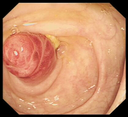
 RSS Feed