Laserna MEJ et.al. Efficacy of Therapy for Eosinophilic Esophagitis in Real-World Practice.
Clinical Gastroenterology & Hepatology. 18(13):2903-2911.e4, 2020 12. ____________________________ Efficacy of Therapy for Eosinophilic Esophagitis in Real-World Practice. Clinical Gastroenterology & Hepatology. 18(13):2903-2911.e4, 2020 12. Authors Laserna-Mendieta EJ; Casabona S; Savarino E; Perello A; Perez-Martinez I et. al. Analysis of data from the EoE CONNECT database-of nearly 600 patients from 11 Spanish centres which was started in 2016 Topical steroids, proton pump inhibitors (PPIs), and dietary interventions are recommended first- and second-line therapies for eosinophilic esophagitis (EoE). We investigated differences in their effectiveness in a real-world, clinical practice cohort of patients with EoE. METHODS: We collected data on the efficacy of different therapies for EoE (ability to induce clinical and histologic remission) from the multicenter EoE CONNECT database-a database of patients with a confirmed diagnosis of EoE in Europe that began in 2016. We obtained data from 589 patients, treated at 11 centers, on sex, age, time of diagnosis, starting date of any therapy, response to therapy, treatment end dates, alternative treatments, and findings from endoscopy. The baseline endoscopy was used for diagnosis of EoE; second endoscopy was performed to evaluate response to first-line therapies. After changes in treatment, generally because lack of efficacy, a last endoscopy was performed. The time elapsed between endoscopies depended on the criteria of attending physicians. Clinical remission was defined by a decrease of more than 50% in Dysphagia Symptom Score; improvement in symptoms by less than 50% from baseline was considered as clinical response. Histologic remission was defined as a peak eosinophil count below 5 eosinophils/hpf. A peak eosinophil count between 5 and 14 eosinophils/hpf was considered histologic response. We identified factors associated with therapy selection and effectiveness using chi2 and multinomial logistic regression analyses RESULTS: Summary of the findings:
Lack of fibrotic features at initial endoscopy was associated with selection of elimination diets over topical steroids as a second-line therapy. The recruiting center was significantly associated with therapy choice; second-line treatment with topical steroids or PPIs were the only variables associated with clinical and histologic remission. CONCLUSIONS: In an analysis of data from a large cohort of patients with EoE in Europe, we found topical steroids to be the most effective at inducing clinical and histologic remission, but PPIs to be the most frequently prescribed. Treatment approaches vary with institution and presence of fibrosis or strictures. PPI was surprisingly effective and seems well worth trying before starting topical steroids Greuter T et.al. Effectiveness and Safety of High- vs Low-Dose Swallowed Topical Steroids for Maintenance Treatment of Eosinophilic Esophagitis: A Multicenter Observational Study.
Clinical Gastroenterology & Hepatology. 2020 Aug 13. ____________________________ Effectiveness and Safety of High- vs Low-Dose Swallowed Topical Steroids for Maintenance Treatment of Eosinophilic Esophagitis: A Multicenter Observational Study. Clinical Gastroenterology & Hepatology. 2020 Aug 13. Authors Greuter T; Godat A; Ringel A; Almonte HS; Schupack D; Mendoza G; McCright-Gill T;, et.al. BACKGROUND & AIMS: Data evaluating efficacy of different doses of swallowed topical corticosteroids (STC) in the long-term management of eosinophilic esophagitis (EoE) are lacking. We assessed long-term effectiveness and safety of different STC doses for adults with EoE after achievement of histological remission. METHODS: We performed a retrospective multicenter study at five EoE referral centers (US and Switzerland). We analyzed data on 82 patients with EoE in histological remission and ongoing STC treatment with therapeutic adherence of >=75% (58 males; mean age at diagnosis, 37.2+/-14.4 years). Patients were followed for a median of 2.2 years (interquartile range [IQR], 1.0-3.8 years). We collected data from 217 follow-up endoscopy visits. The primary endpoint was time to histological relapse. RESULTS: Histological relapse occurred in 67% of patients. Relapse rates were comparable in patients taking low dose (<=0.5 mg per day, n = 58) and high dose STC (>0.5 mg per day, n = 24) with 72 vs 54% (ns). However, histological relapse occurred significantly earlier with low dose STC (1.0 vs 1.8 years, P = .030). There was no difference regarding rates of and time to stricture formation for low vs high dose STC. Esophageal candidiasis was observed in 6% of patients (5% for low dose, 8% for high dose, ns). No dysplasia or mucosal atrophy was detected. CONCLUSION: Histological relapse frequently occurs in EoE despite ongoing STC treatment regardless of STC doses. However, relapse develops later in patients on high dose STC without an increase in side-effects. Doses higher than 0.5 mg/day may be considered for EoE maintenance treatment, but advantage over lower doses appears to be small. Copyright © 2020 AGA Institute. Published by Elsevier Inc. All rights reserved. Presumably this was patients who had attained remission with topical steroids? Remember that about 1/3 of patients don’t respond. Of course, the potential benefits of systemic glucocorticosteroids in EoE patients that are refractory to topical glucocorticosteroids are currently unknown. Ding W et.al. Endoscopic retrograde appendicitis therapy (ERAT) for management of acute appendicitis. Surgical Endoscopy. 2021 May 13.
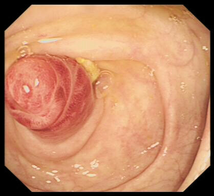
____________________________ Endoscopic retrograde appendicitis therapy (ERAT) for management of acute appendicitis. Surgical Endoscopy. 2021 May 13. Authors Ding W; Du Z; Zhou X BACKGROUND: This study investigated the feasibility of endoscopic retrograde appendicitis therapy (ERAT) for the treatment of acute appendicitis. METHODS: There were 210 patients included who were admitted to our hospital from January 2017 to October 2019 with a diagnosis of acute appendicitis. According to the method of treatment, patients were stratified into the ERAT group, laparoscopic appendectomy (LA) group, or open appendectomy (OA) group for comparison of perioperative information extracted from the medical records of the patients. RESULTS: The operations were successfully completed in all patients. The length of operation in the endoscopy group (median: 48 min, range: 34-78 min) was significantly shorter compared to the Laparoscopic group (median: 67 min, range: 47-90 min) or open surgery group (median: 85 min, range: 58-120 min). Postoperatively, the length of stay, the amount of time spent bedridden following surgery, surgery-related complications, and in-patient expenses were all significantly less in the Endoscopic than in pts managed surgically (all p < 0.05). Moreover, the recurrence rate of appendicitis after Endoscopic treatment was 1: 30 (2.86%) during the first 6 months of postoperative follow-up. 13 patients who underwent the Endoscopic treatment developed an appendicular abscess. These were all successfully treated colonoscopically incising the most protruding or fluctuating place around the appendix opening without procedure-related complications during the follow-up period. CONCLUSION: Endoscopic treatment is a safe and effective endoscopic treatment method for acute appendicitis and abscesses of the appendix. The advantages include reduced trauma, faster recovery times, and lower costs in comparison with either surgical procedures. ERAT with internal incision and drainage can be safely performed with immediate effect, especially in patients with acute uncomplicated appendicitis accompanied by either fecal stones or stenosis of the appendix cavity, or an abscess within the appendix cavity. To remind you, the idea that we may be able to treat appendicitis goes back to 2012
Becq A et.al. ERCP within 6 or 12 h for acute cholangitis: a propensity score-matched analysis. Surgical Endoscopy. 2021 May 11.
____________________________ ERCP within 6 or 12 h for acute cholangitis: a propensity score-matched analysis. Surgical Endoscopy. 2021 May 11. Authors Becq A; Chandnani M; Bartley A; Nuzzo A; Bilal M; Bharadwaj S; Cohen J; et.al. BACKGROUND: The optimal timing of ERCP for patients with acute cholangitis remains controversial. The aim of our study was to determine if ERCP performed within 6 or 12 h of presentation was associated with improved clinical outcomes. METHODS: Medical records for all patients with acute cholangitis who underwent ERCP at our institution between 2009 and 2018 were reviewed. Outcomes were compared between those who underwent ERCP within or after 12 h using propensity score framework. Our primary outcome was length of hospitalization. Secondary outcomes included in-hospital mortality, adverse events, ERCP failure, length of ICU stay, organ failure, recurrent cholangitis, and 30-day readmission. In secondary analysis, outcomes for ERCP done within or after 6 h were also compared. RESULTS: During study period, 487 patients with cholangitis were identified, of whom 147 (30% of all patients) had ERCP within 12 h of presentation. Using propensity score matching, we selected 145 pairs of patients with similar characteristics.
Of course, most tertiary referral centres are unable to offer emergency ERCP in the middle of the night. It would have been interesting to learn about the outcomes in comparing ERCP within 12 hours vs within 24 hours. My own take-home message is that any patient with evidence of septic shock need ERCP within 24 hours and perhaps even within 12 hours (which of course means the next day). As you know, acute cholangitis is a medical emergency linked with a 2-5% mortality rate and ERCP can be life-saving
Ashkar MH et.al. Increased Risk of Advanced Colonic Adenomas and Timing of Surveillance Colonoscopy Following Solid Organ Transplantation.
Digestive Diseases & Sciences. 2021 May 10. ____________________________ Increased Risk of Advanced Colonic Adenomas and Timing of Surveillance Colonoscopy Following Solid Organ Transplantation. Digestive Diseases & Sciences. 2021 May 10. Authors Ashkar MH; Chen J; Shy C; Crippin JS; Chen CH; Sayuk GS; Davidson NO BACKGROUND: Detection and removal of colonic adenomatous polyps decreases colorectal cancer (CRC) development, particularly with more or larger polyps or polyps with advanced villous/dysplastic histology. Immunosuppression following solid organ transplantation may accelerate adenomas development and progression compared to average-risk population but the benefit of earlier colonoscopic surveillance is unclear. AIMS: Study the impact of maintenance immunosuppression post-transplantation on developmental timing, multiplicity and pathological features of adenomas, by measuring incidence of advanced adenomas (villous histology, size >= 10 mm, >= 3 polyps, presence of dysplasia) post-transplantation and the incidence of newly diagnosed CRC compared to average-risk age-matched population. METHODS: Single-center retrospective cohort study of transplantation recipients. RESULTS: 295 transplantation recipients were included and were compared with 291 age-matched average-risk controls. The mean interval between screening and surveillance colonoscopies between transplantation and control groups was 6.3 years vs 5.9 years (p = 0.13). Post-transplantation maintenance immunosuppression mean duration averaged 59.9 months at surveillance colonoscopy. On surveillance, transplantation recipients had more large adenomas (10 mm or larger) compared to matched controls (9.2% vs. 3.8%, p = 0.034; adjusted OR 2.38; 95% CI 1.07-5.30). CONCLUSION: transplantation recipients appear at higher risk for developing advanced adenomas, suggesting that earlier surveillance should be considered. Does immunosuppression also suppress our bodies ability to keep rouge DNA in check? Or does this study simply show that if you are on a transplant list, attending for screening colonoscopy is a low priority? Chandrasekhara V et.al. Predicting the need for step-up therapy after EUS-guided drainage of pancreatic fluid collections with lumen-apposing metal stents.
Clinical Gastroenterology & Hepatology. 2021 May 06. ____________________________ Predicting the need for step-up therapy after EUS-guided drainage of pancreatic fluid collections with lumen-apposing metal stents. Clinical Gastroenterology & Hepatology. 2021 May 06. Authors Chandrasekhara V; Elhanafi S; Storm AC; Takahashi N, et.al. BACKGROUND & AIMS: A significant proportion of individuals with pancreatic fluid collections (PFCs) require step-up therapy after endoscopic drainage with lumen-apposing metal stents (LAMS). The aim of this study is to identify factors associated with PFCs that require step-up therapy. METHODS: A retrospective cohort study of patients undergoing EUS-guided drainage of PFCs with LAMS from 4/2014 to 10/2019 at a single center was performed.
RESULTS: 136 patients who had their collections drained with a stent, were included in the final study cohort, of whom 69 (50.7%) required step-up therapy. Independent predictors of step-up therapy included:
CONCLUSIONS: Half of all patients with PFCs drained with LAMS required step-up therapy, most commonly DEN. Individuals with PFCs >10 cm in size, paracolic extension, or >30% solid necrosis are more likely to require step-up therapy and should be considered for early endoscopic re-intervention. . Messallam AA et.al. Endoscopic Necrosectomy With and Without Hydrogen Peroxide for Walled-off Pancreatic Necrosis: A Multicenter Comparative Study.
American Journal of Gastroenterology. 116(4):700-709, 2021 Apr. ____________________________ Endoscopic Necrosectomy With and Without Hydrogen Peroxide for Walled-off Pancreatic Necrosis: A Multicenter Comparative Study. American Journal of Gastroenterology. 116(4):700-709, 2021 Apr. Authors Messallam AA; Adler DG; Shah RJ; Nieto JM, et.al. INTRODUCTION: Endoscopic necrosectomy has emerged as the preferred treatment modality for walled-off pancreatic necrosis. This study was designed to evaluate the safety and efficacy of direct endoscopic necrosectomy with and without hydrogen peroxide (H2O2) lavage. METHODS: Retrospective chart reviews were performed for all patients undergoing endoscopic transmural management of walled-off pancreatic necrosis at 9 major medical centers from November 2011 to August 2018. Clinical success was defined as the resolution of the collection by imaging within 6 months, without requiring non-endoscopic procedures or surgery. RESULTS: Of 293 patients, 204 patients met the inclusion criteria. Technical and clinical success rates were 100% (204/204) and 81% (166/189), respectively. For patients, 122 (59.8%) patients had at least one hydrogen peroxide necrosectomy and 82 (40.2%) patients had standard endoscopic necrosectomy.
Shiroma S et.al. Timing of bleeding and thromboembolism associated with endoscopic submucosal dissection for gastric cancer in Japan.
Journal of Gastroenterology & Hepatology. 2021 May 07 ____________________________ Timing of bleeding and thromboembolism associated with endoscopic submucosal dissection for gastric cancer in Japan. Journal of Gastroenterology & Hepatology. 2021 May 07. Authors Shiroma S; Hatta W; Tsuji Y; Yoshio T; Yabuuchi Y, et.al. OBJECTIVE: This study aimed to reveal the timing of bleeding and thromboembolism associated with endoscopic submucosal dissection (ESD) for early gastric cancer (EGC). METHODS: We retrospectively reviewed 10,320 patients who underwent ESD for EGC during November 2013-October 2016. We evaluated overall bleeding rates and their inter-group differences. Factors associated with early/late (cut-off 5 days) bleeding and thromboembolism frequency and its association with the intake of antithrombotic (AT) agents were investigated. RESULTS: Overall, the post-ESD bleeding rate was 4.7% (489/10,320);
Of course we already know that the risk of bleeding after gastric EMR and gastric ESD is the same. I will be using this data when I consent patients for a gastric resection of any lesion. Yasuda T et.al. Benefits of linked color imaging for recognition of early differentiated-type gastric cancer: in comparison with indigo carmine contrast method and blue laser imaging.
Surgical Endoscopy. 35(6):2750-2758, 2021 Jun. ____________________________ Benefits of linked color imaging for recognition of early differentiated-type gastric cancer: in comparison with indigo carmine contrast method and blue laser imaging. Surgical Endoscopy. 35(6):2750-2758, 2021 Jun. Authors Yasuda T; Yagi N; Omatsu T; Hayashi S; Nakahata Y, et/al. BACKGROUND AND AIM: Linked colour imaging is proprietary Fujifilm technology which enhances slight differences in mucosal color. However, whether LCI is more useful than other kinds of image-enhanced endoscopy (IEE) in recognizing early gastric cancer remains unclear. This study aimed to evaluate LCI efficacy compared with the indigo carmine contrast method (IC), and blue laser imaging-bright (BLI-brt) in early differentiated-type gastric cancer recognition. METHODS: We retrospectively analyzed early differentiated-type gastric cancer, which were examined by all four imaging techniques (white light imaging, IC, LCI, BLI-brt) at Asahi University Hospital from June 2014 to November 2018. Both subjective evaluation (using ranking score: RS) and objective evaluation (using color difference score: CDS) were adopted to quantify early differentiated-type gastric cancer recognition. RESULTS: During this period, 87 EGC lesions were enrolled in this study. Both RS and CDS of LCI were significantly higher (p < 0.01) than those of IC and BLI-brt. Both RS and CDS of BLI-brt had no significant difference compared with those of IC. Subgroup analysis indicated that Linked colour imaging may be better than Indigo carmine dye particularly in finding flat or depressed lesions. CONCLUSIONS: LCI appears to be more beneficial for the recognition of early differentiated-type gastric cancer in endoscopic screenings than IC and BLI-brt from the middle to distant view. Kim JW et.al. Narrowband imaging with near-focus magnification for discriminating the gastric tumor margin before endoscopic resection: A prospective randomized multicenter trial.
Journal of Gastroenterology & Hepatology. 35(11):1930-1937, 2020 Nov. ____________________________ Narrowband imaging with near-focus magnification for discriminating the gastric tumor margin before endoscopic resection: A prospective randomized multicenter trial. Journal of Gastroenterology & Hepatology. 35(11):1930-1937, 2020 Nov. Authors Kim JW; Jung Y; Jang JY; Kim GH; Bang BW; Park JC; Choi HS, et.al. Prospective randomized controlled trial was conducted at seven teaching hospitals in South Korea BACKGROUND AND AIM: This study investigated the usefulness of near-focus narrowband imaging (NF-NBI) for determining gastric tumor margins compared with indigo carmine chromoendoscopy (ICC) before endoscopic submucosal dissection (ESD). METHODS: This prospective randomized controlled trial was conducted at seven teaching hospitals in Korea. Patients with gastric adenoma or differentiated adenocarcinoma undergoing ESD were enrolled and randomly assigned to the NF-NBI or ICC group. A marking dot was placed on the most proximal margin of the tumor before ESD. The primary endpoint was delineation accuracy, which was defined as presence of marking dots within 1 mm of the tumor margin under microscopic observation. RESULTS: A total of 200 patients had ‘Near Focus NBI’ and 195 patients had indigo carmine dye to delineate the margin of the EGC’s. The delineation Accuracy rate was 85% in the ‘Near Focus NBI’ group and 80.0% in the ‘indigo carmine dye spray’ group (P = 0.44). However, the distance from the marking dot to the margin of the tumor was significantly shorter in the ‘Near Focus NBI’ group than in the ‘indigo carmine dye spray’ group (0.8 +/- 0.8 vs 1.2 +/- 1.3 mm, P < 0.01). Even after adjustment of other clinicopathological factors that are associated with difficulty of tumor delineation, ‘Near Focus NBI’ did not show significant association with accurate delineation (odds ratio of 0.86, P = 0.60). CONCLUSIONS: The authors concluded that Near Focus and NBI was not significantly better than Indigo Carmine spray to accurately delineating gastric tumors Surek A et.al. Risk factors affecting failure of colonoscopic detorsion for sigmoid colon volvulus: a single center experience.
International Journal of Colorectal Disease. 36(6):1221-1229, 2021 Jun. ____________________________ Risk factors affecting failure of colonoscopic detorsion for sigmoid colon volvulus: a single center experience. International Journal of Colorectal Disease. 36(6):1221-1229, 2021 Jun. Authors Surek A; Akarsu C; Gemici E; Ferahman S; Dural AC; Bozkurt MA, et.al. PURPOSE: Colonoscopic detorsion (CD) is the first treatment option for uncomplicated sigmoid volvulus (SV). We aim to examine the factors affecting the failure of CD. METHODS: The files of patients, treated after diagnosis of SV between January 2015 and September 2020, were retrospectively reviewed. Patients' demographic data, comorbidities, endoscopy reports, and surgical and other treatments were recorded. Patients were divided into two groups, as the successful CD group and unsuccessful CD group. The data were compared between the groups, and multivariate analysis of statistically significant variables was performed. RESULTS: There were 21 (30%) patients had a failed sigmoidoscopic detorsion and 52 patients underwent a successful procedure. [This is greater than the 95% success rate reported in previous studies but of course, the chance of success must be very operator dependent]. The unsuccessful CD rate was found to be 28.76%; this is likely a function of. Risk factors for failure included;
values as statistically significant. In the multivariate analysis, previous abdominal surgery and cecum diameter over 10 cm were seen as predictive factors for failure of CD (p=0.049, OR=0.103, and p = 0.028, OR=10.540, respectively). CONCLUSIONS: CD failure rate was significantly associated with previous abdominal surgery and a cecum diameter over 10 cm. We found that patients with these factors will tend to need more emergency surgery. Not for the first time in medicine do we find that the patients in the greatest need of a successful outcome of a medical intervention, is precisely the group who is the least likely to benefit ! Clark G et.al. Transition to quantitative faecal immunochemical testing from guaiac faecal occult blood testing in a fully rolled-out population-based national bowel screening programme.
Gut. 70(1):106-113, 2021 Jan. ____________________________ Transition to quantitative faecal immunochemical testing from guaiac faecal occult blood testing in a fully rolled-out population-based national bowel screening programme. Gut. 70(1):106-113, 2021 Jan. Authors Clark G; Strachan JA; Carey FA; Godfrey T; Irvine A; McPherson A; Brand J; Anderson AS, et.al. OBJECTIVE: Faecal immunochemical tests (FIT) are replacing guaiac faecal occult blood tests (FOBT) in colorectal cancer (CRC) screening. Data from the first year of FIT screening were compared with those from FOBT screening and assumptions based on a pilot evaluation of FIT. DESIGN: Data on uptake, positivity, positive predictive value (PPV) for CRC and higher-risk adenoma from participants in the first year of the FIT-based Scottish Bowel Screening Programme (n=919 665), with a threshold of 80 microg Hb/g faeces, were compared with those from the penultimate year of the FOBT-based programme (n=862 165) and those from the FIT evaluation (n=66 225). RESULTS: Overall
CONCLUSION: Transition to FIT from FOBT produced higher uptake and positivity with lower PPV for CRC and higher PPV for adenoma. The FIT pilot evaluation underestimated uptake and positivity. Introducing FIT at the same threshold as the evaluation caused a 67.2% increase in colonoscopy demand instead of a predicted 10% The whole of the UK has moved from guaiac based FOB testing to faecal immunochemical FIT test. This study only pertains to the Scottish experience but I see no reason why their findings would be different to the UK experience as a whole. I remember being told that the best thing about FIT testing is that you can change the sensitivity of the test so that my colonoscopy service doesn’t become overwhelmed. Well that was never going to happen was it? That dial is welded into place and nothing short of a declaration in the house of Parliament would move it Gachabayov M et.al. Performance evaluation of stool DNA methylation tests in colorectal cancer screening: a systematic review and meta-analysis.
Colorectal Disease. 23(5):1030-1042, 2021 05. ____________________________ Performance evaluation of stool DNA methylation tests in colorectal cancer screening: a systematic review and meta-analysis. Colorectal Disease. 23(5):1030-1042, 2021 05. Authors Gachabayov M; Lebovics E; Rojas A; Felsenreich DM; Latifi R; Bergamaschi R AIM: There is not sufficient evidence about whether stool DNA methylation tests allow prioritizing patients to colonoscopy. Due to the COVID-19 pandemic, there will be a wait-list for rescheduling colonoscopies once the mitigation is lifted. The aim of this meta-analysis was to evaluate the accuracy of stool DNA methylation tests in detecting colorectal cancer. METHODS: 46 studies including a total of 16,000 patients were included in this meta-analysis. The PubMed, Cochrane Library and MEDLINE via Ovid were searched. Studies reporting the accuracy (Sackett phase 2 or 3) of stool DNA methylation tests to detect sporadic colorectal cancer were included. The DerSimonian-Laird method with random-effects model was utilized for meta-analysis. RESULTS: 46 studies totaling 16 149 patients were included in the meta-analysis. The pooled sensitivity and specificity of all single genes and combinations was 62.7% (57.7%, 67.4%) and 91% (89.5%, 92.2%), respectively. Combinations of genes provided higher sensitivity compared to single genes (80.8% [75.1%, 85.4%] vs. 57.8% [52.3%, 63.1%]) with no significant decrease in specificity (87.8% [84.1%, 90.7%] vs. 92.1% [90.4%, 93.5%]).
CONCLUSIONS: Stool DNA methylation tests have high specificity (92%) with relatively lower sensitivity (81%). Combining genes increases sensitivity compared to single gene tests. The single most accurate gene is SDC2, which should be considered for further research. Clearly testing stool for DNA methylation is almost but not quite ready for prime time ! Forbes N et.al. Association Between Endoscopist Annual Procedure Volume and Colonoscopy Quality: Systematic Review and Meta-analysis.
Clinical Gastroenterology & Hepatology. 18(10):2192-2208.e12, 2020 09. ____________________________ Association Between Endoscopist Annual Procedure Volume and Colonoscopy Quality: Systematic Review and Meta-analysis. Clinical Gastroenterology & Hepatology. 18(10):2192-2208.e12, 2020 09. Authors Forbes N; Boyne DJ; Mazurek MS; Hilsden RJ; Sutherland RL, et.al. A systematic review of 27 studies of 11,000 000 colonoscopies looking at flight hours vs quality BACKGROUND & AIMS: In addition to monitoring adverse events (AEs) and post-colonoscopy colorectal cancers (PCCRC), indicators for assessing colonoscopy quality include adenoma detection rate (ADR) and cecal intubation rate (CIR). It is unclear whether there is an association between annual colonoscopy volume and ADR, CIR, AEs, or PCCRC. METHODS: We searched publication databases through March 2019 for studies assessing the relationship between annual colonoscopy volume and outcomes, including ADR, CIR, AEs, or PCCRC. Pooled odds ratios (ORs) were calculated using DerSimonian and Laird random effects models. Sensitivity analyses were performed to assess for potential methodological or clinical factors associated with outcomes. RESULTS: We performed a systematic review of 9235 initial citations, generating 27 retained studies comprising 11,276,244 colonoscopies.
CONCLUSIONS: In a systematic review and meta-analysis, we found higher annual colonoscopy volumes to correlate with higher CIR, but not with ADR or PCCRC. Trends toward fewer AEs were associated with higher annual colonoscopy volumes. There are few data available from endoscopists who perform fewer than 100 annual colonoscopies. Studies are needed on extremes in performance volumes to more clearly elucidate associations between colonoscopy volumes and outcomes. Of course, hardly any studies included colonoscopists who did fewer than 100 procedures a year. It looks like endoscopists who only do a small number of colonoscopies each year don’t readily volunteer their data. Is there a threshold effect in that if you do a certain number of procedures each year, your ‘performance’ levels off and you don’t find any more adenomas ? Fruehauf H et.al. Evaluation of Acute Mountain Sickness by Unsedated Transnasal Esophagogastroduodenoscopy at High Altitude.
Clinical Gastroenterology & Hepatology. 18(10):2218-2225.e2, 2020 09. ____________________________ Evaluation of Acute Mountain Sickness by Unsedated Transnasal Esophagogastroduodenoscopy at High Altitude. Clinical Gastroenterology & Hepatology. 18(10):2218-2225.e2, 2020 09. Authors Fruehauf H; Vavricka SR; Lutz TA; Gassmann M; Wojtal KA; Erb A, et.al. A prospective study, 25 Swiss mountaineers BACKGROUND & AIMS: It is not clear how rapid ascent to a high altitude causes the gastrointestinal symptoms of acute mountain sickness (AMS). We assessed the incidence of endoscopic lesions in the upper gastrointestinal tract in healthy mountaineers after a rapid ascent to high altitude, their association with symptoms, and their pathogenic mechanisms. METHODS: In a prospective study, 25 mountaineers (10 women; mean age, 45 underwent unsedated, transnasal esophagogastroduodenoscopy at ground level and then after 4 days in at a high altitude laboratory in the Alps (at 4559 meters). Symptoms were assessed using validated instruments for AMS (the acute mountain sickness score and the Lake Louise scoring system) and visual analogue scales (scale, 0-100). Levels of messenger RNAs (mRNAs) in duodenal biopsy specimens were measured by quantitative polymerase chain reaction. RESULTS: The follow-up endoscopy at high altitude was performed in 19 of 25 patients on day 2 and in 23 of 25 patients on day 4.
I’m looking forward to a study of colonoscopy at the top of Mount Everest. Goverde A et.al. Yield of Lynch Syndrome Surveillance for Patients With Pathogenic Variants in DNA Mismatch Repair Genes.
Clinical Gastroenterology & Hepatology. 18(5):1112-1120.e1, 2020 05. ____________________________ Yield of Lynch Syndrome Surveillance for Patients With Pathogenic Variants in DNA Mismatch Repair Genes. Clinical Gastroenterology & Hepatology. 18(5):1112-1120.e1, 2020 05. Authors Goverde A; Eikenboom EL; Viskil EL; Bruno MJ et.al. A Retrospective analysis of surveillance data from a single centre in the Netherlands (Rotterdam) BACKGROUND & AIMS: Patients with Lynch syndrome are offered the same colorectal cancer (CRC) surveillance programs (colonoscopy every 2 years), regardless of the pathogenic DNA mismatch repair gene variant the patient carries. We aimed to assess the yield of surveillance for patients with these variants in MLH1, MSH2, MSH6, and PMS2. METHODS: We analyzed data on colonoscopy surveillance, including histopathology analysis, from all patients diagnosed with Lynch syndrome (n = 264 with Lynch Syndrome) at a single center. We compared the development of (advanced) adenomas and CRC among patients with pathogenic variants in the DNA mismatch repair genes MLH1 (n = 55), MSH2 (n = 44), MSH6 (n = 143), or PMS2 (n = 22) over 1836 years of follow-up (median follow-up of 6 years per patient). RESULTS: At first colonoscopy, CRC was found in 8 patients. During 916 surveillance colonoscopies, CRC was found in 9 patients and 6 patients died during follow up (5 from some cancer).. No CRC was found in patients with variants in MSH6 or PMS2 over the entire follow-up period. There were no significant differences in the number of colonoscopies with adenomas or advanced adenomas among the groups. The median time of adenoma development was 3 years (IQR, 2-6 years). There were no significant differences in time to development of adenoma.
CONCLUSIONS: No CRC was found during follow-up of patients with Lynch syndrome carrying pathogenic variants in MSH6; advanced neoplasia developed over shorter follow-up time periods in patients with pathogenic variants in MLH1 or MSH2. The colonoscopy interval for patients with pathogenic variants in MSH6 might be increased to 3 years from the regular 2-year interval. Mutations in the MSH6 mismatch repair is different! Lamba M et.al. Associations Between Mutations in MSH6 and PMS2 and Risk of Surveillance-detected Colorectal Cancer.
Clinical Gastroenterology & Hepatology. 18(12):2768-2774, 2020 11. ____________________________ Associations Between Mutations in MSH6 and PMS2 and Risk of Surveillance-detected Colorectal Cancer. Clinical Gastroenterology & Hepatology. 18(12):2768-2774, 2020 11. Authors Lamba M; Wakeman C; Ebel R; Hamilton S, et.al. Prospective study of 381 persons with Lynch syndrome in New Zealand BACKGROUND & AIMS: Lynch syndrome is the most common inherited cause of colorectal cancer (CRC). Contemporary and mutation-specific estimates of CRC-risk in patients undergoing colonoscopy would optimize surveillance strategies. We performed a prospective national cohort study, using data from New Zealand, to assess overall and mutation-specific risk of CRC in patients with Lynch syndrome undergoing surveillance. METHODS: We performed a prospective study of 381 persons with Lynch syndrome in New Zealand (98 with Lynch-syndrome associated variants in MLH1, 159 in MSH2, 103 in MSH6, and 21 in PMS2). Participants were offered annual colonoscopy starting at age 25 y, and those who underwent 2 or more colonoscopies before December 31, 2017 were included in the final analysis. Patients with previous colonic resection, history of CRC or diagnosis of CRC at index colonoscopy were excluded. RESULTS: Study participants underwent 2061 colonoscopies during a median of 4.4 year follow up 2296 person-y; the median observation-period was 4.43 y and mean-age at enrollment was 43 y.
Age-adjusted CRC-risk in patients with variants in MSH6 was lower than in MLH1 (hazard ratio, 0.2; 95% CI, 0.04-0.94; P = .02). Of patients with CRC, 33% had an adenomatous polyp resected from same segment in which a colorectal tumor later developed. CONCLUSIONS: The risk of CRC in patients with Lynch syndrome-associated mutations in MSH6 or PMS2 was significantly lower than in patients with mutations in MLH1. Incomplete adenomatous polyp resection might be responsible for one third of surveillance-detected CRCs. Should we relax a little on surveillance intensity in patients with Lynch syndrome and the MSH6 variant? Alternatively, as the risk of cancer was close to 20%, should patients with the MLH1 or the MSH2 mutations be offered total colectomy and only patients with MSH6 mutations be offered surgery? The again nobody died from their bowel cancer. And patients with Lynch syndrome have increased risk of small bowel cancer, pancreatic cancer, renal cancer, biliary tract cancer, brain cancer, prostate cancer and breast cancer ! We can’t remove or surveil most of these areas Omidvar AH et.al. The optimal age to stop endoscopic surveillance of Barrett's esophagus patients based on sex and comorbidity: a comparative cost-effectiveness analysis.
Gastroenterology. 2021 May 08. ____________________________ The optimal age to stop endoscopic surveillance of Barrett's esophagus patients based on sex and comorbidity: a comparative cost-effectiveness analysis. Gastroenterology. 2021 May 08. Authors Omidvari AH; Hazelton WD; Lauren BN, et.al. BACKGROUND & AIMS: Current guidelines recommend surveillance for non-dysplastic Barrett's esophagus (NDBE) patients but do not include a recommended age for discontinuing surveillance. This study aimed to determine the optimal age for last surveillance of NDBE patients stratified by sex and level of comorbidity. METHODS: We used 3 independently developed models to simulate patients with stable Barrett’s, varying in age, sex, and comorbidity level (no, mild, moderate, severe). All patients had received regular surveillance until their current age. We calculated incremental costs and quality-adjusted life-years (QALYs) gained from one additional endoscopic surveillance at the current age versus not performing surveillance at that age. We determined the optimal age to end surveillance as the age at which incremental cost-effectiveness ratio (ICER) of one more surveillance was just below the willingness-to-pay threshold of $100,000/QALY. RESULTS: The benefit of having one more surveillance endoscopy strongly depended on age, sex and comorbidity. Carrying out 1000 Barrett’s surveillance gastroscopies in 80 yr old men with previously stable Barrett’s but with a severe comorbidity, provided 4 more quality-adjusted life-years (QALYs) at a cost of $1,2 million, while for women with severe comorbidity the benefit at that age was 7 QALYs at a cost of $1.3 million. The authors concluded that the best time to stop was:
CONCLUSIONS: Our comparative modelling analysis illustrates the importance of considering comorbidity status and sex when deciding upon the age to discontinue surveillance in patients with NDBE. My generation has completely failed to pay any attention the cost, resource implications and climate impact of surveillance. We subject our patients to uncomfortable surveillance from which they are very unlikely to benefit whilst squandering money and resources which could be used to provide these patients with health care which provides better value for the elderly. Hip replacements, cataract surgery or perhaps better community care or whatever. Surely, it’s not unreasonable to conclude that there comes a time in a patients life when surveillance is no longer appropriate and spending the money on other types of care is more appropriate?
0 Comments
Leave a Reply. |
Archives
June 2022
Categories
All
|


 RSS Feed