Friends of Endoscopy
  • Home
  • Quiz Cases
  • Endoscopy Long Cases
  • Short Teaching Clips
  • Blackboard teaching
  • Podcasts
  • Core Reading
    • Basic concepts core reading
    • Gastroscopy core reading
    • Colonoscopy core reading
    • QA core reading
  • About
    • Our sponsors
  • Home
  • Quiz Cases
  • Endoscopy Long Cases
  • Short Teaching Clips
  • Blackboard teaching
  • Podcasts
  • Core Reading
    • Basic concepts core reading
    • Gastroscopy core reading
    • Colonoscopy core reading
    • QA core reading
  • About
    • Our sponsors
Search
Picture
​

Friends of Endoscopy is all about pattern recognition.  See it today and recognise it tomorrow!   Learn from a New Case on most weekdays !!! 
Become a Better Endoscopist ! 

A common oesophageal finding?

15/2/2021

 
Picture
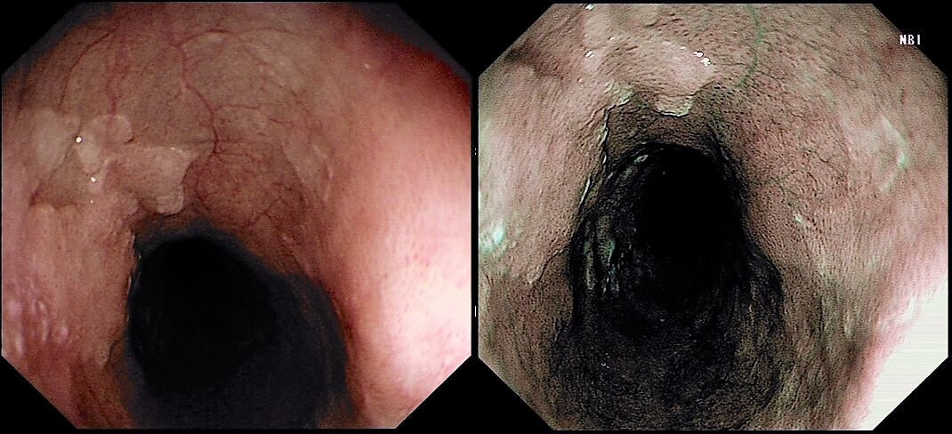
This was found in the distal oesophagus of a patient with dyspepsia. You take some samples. 
WHAT WILL YOU TELL THE PATIENT?
■ This is a benign finding
Yes it is but do you know what it is?
■ We'll see what biopsies show
It's' a spot diagnosis!
■ This may well need EMR
Very unlikely unless those IPCL's look odd
■ Staging investigations are likely next
Can't imagine!
explanation
This may be a good example of 'we don't see' because we don't know what it is. Intuitively, we all blank this because it's a benign finding. But what is it?  It's actually a patch of squamous hyperkeratosis with some underlying basal cell hyperplasia. The normal squamous basal cell layer is 1-4 cells thick but, in reflux oesophagitis thic increase up to 75% of the epithelial thickness. However, many cases of reflux oesophagitis does not exhibit basal cell hyperplasia and conversely, there are cases which will not be due to reflux disease as in this case!  

In rodents (!), the finding has been linked with vitamin deficiencies and zinc deficiency.  Of course, squamous hyperkeratosis in the mouth is associated with squamous neoplasia [ J Clin Pathol 2005;58:1325–1327 ] and for this reason, I always take some samples.  However, I am yet to find a single case of dysplasia within a patch of oesophageal squamous hyperkeratosis !!! 
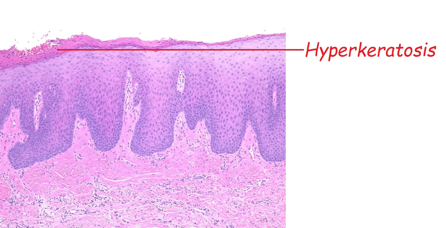
Picture

    Categories

    All
    Barrett's
    Cancer
    Cancer Syndromes
    Colitis
    Colorectum
    Corrosive Ingestion
    Crypt Pattern
    Difficulty: Hard
    Difficulty: Moderate
    Difficulty: Very Hard
    Duodenum
    Eosinophilic Oesophagitis
    EUS
    Foveolar Metaplasia
    Gastric
    Gastroscopy
    GI Bleeding
    Histology
    HPB
    Ileum
    Immunosuppression
    Infection
    Local Recurrence Of Barrett's Ca.
    Lymphoma
    Mixed Polyp
    Mucosal Prolapse Syndrome
    NET
    Non-lifting Sign
    Oesophagus
    Opinion Piece!
    Pharynx
    Polyp
    Polypectomy
    The Basics
    TSA

  • Home
  • Quiz Cases
  • Endoscopy Long Cases
  • Short Teaching Clips
  • Blackboard teaching
  • Podcasts
  • Core Reading
    • Basic concepts core reading
    • Gastroscopy core reading
    • Colonoscopy core reading
    • QA core reading
  • About
    • Our sponsors