Friends of Endoscopy
  • Home
  • Quiz Cases
  • Endoscopy Long Cases
  • Short Teaching Clips
  • Blackboard teaching
  • Podcasts
  • Core Reading
    • Basic concepts core reading
    • Gastroscopy core reading
    • Colonoscopy core reading
    • QA core reading
  • About
    • Our sponsors
  • Home
  • Quiz Cases
  • Endoscopy Long Cases
  • Short Teaching Clips
  • Blackboard teaching
  • Podcasts
  • Core Reading
    • Basic concepts core reading
    • Gastroscopy core reading
    • Colonoscopy core reading
    • QA core reading
  • About
    • Our sponsors
Search
Picture
​

Friends of Endoscopy is all about pattern recognition.  See it today and recognise it tomorrow!   Learn from a New Case on most weekdays !!! 
Become a Better Endoscopist ! 

An incidental oesophageal finding

19/3/2021

Comments

 
Picture
Picture
Picture
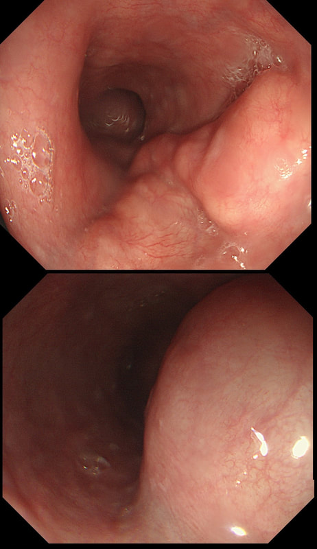
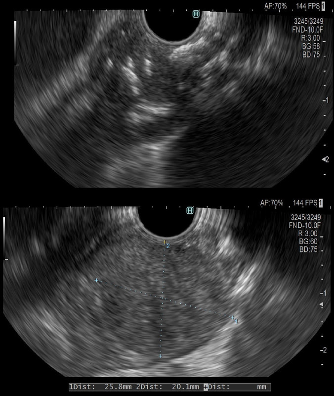
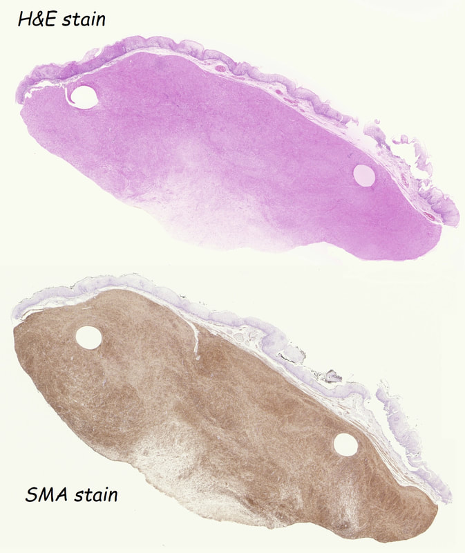
This was an incidental finding in the mid-oesophagus. Subsequently an EUS is organised and the lesion is ultimately removed (histology in the last image)
WHAT IS THE DIAGNOSIS?
■ Neuroendocrine tumour
I've never actually seen an oesophageal NET
■ Granular cell tumour
But it isn't a small white submucosal disc?!
■ Leiomyoma
A shrewd guess!
■ GIST
These are VERY rare in the oesophagus (but common in stomach)
■ Melanoma
Usually look brownish endoscopically!
explanation
Leiomyomas are the most common submucosal lesion found in the oesophagus. No idea why they are so rare in the stomach, where GIST's are the most common submucosal lesion. Correspondingly GIST's are very rare in the oesophagus.

These usually arise from the circular muscle propria layer. On CT they are often calcified and there are of course no nearby nodes to suggest malignant disease.  Indeed, malignant transformation is extremely rare and for this reason many centres would ignore a small submucosal lesion such as this, when found in an elderly person. Larger (around 5cm usually) and symptomatic lesions (which are more likely to be leiomyosarcomas) are enucleated surgically. "- They shell out like peas" according to one of our UGI surgeons.

However, our local surgeons are also careful to point out that resection usually result in some damage to the muscle propria layer, which require repair as otherwise a diverticulum may develop.

With endoscopists spending more time in the 'third oesophageal space' (i.e. the submucosal space), doing ESD's and POEM, the suggestion that we should also remove leiomyomas endoscopically has arisen. May seem like a good idea but I would advocate caution for the simple reason that endoscopically, we are unable to repair the defect we cause in the muscle propria layer ... 

Oh, how about that 'SMA stain' used in the histology.  This is 'Smooth Muscle Actin' stain which does exactly what it says on the tin ... 
Comments

    Categories

    All
    Barrett's
    Cancer
    Cancer Syndromes
    Colitis
    Colorectum
    Corrosive Ingestion
    Crypt Pattern
    Difficulty: Hard
    Difficulty: Moderate
    Difficulty: Very Hard
    Duodenum
    Eosinophilic Oesophagitis
    EUS
    Foveolar Metaplasia
    Gastric
    Gastroscopy
    GI Bleeding
    Histology
    HPB
    Ileum
    Immunosuppression
    Infection
    Local Recurrence Of Barrett's Ca.
    Lymphoma
    Mixed Polyp
    Mucosal Prolapse Syndrome
    NET
    Non-lifting Sign
    Oesophagus
    Opinion Piece!
    Pharynx
    Polyp
    Polypectomy
    The Basics
    TSA

  • Home
  • Quiz Cases
  • Endoscopy Long Cases
  • Short Teaching Clips
  • Blackboard teaching
  • Podcasts
  • Core Reading
    • Basic concepts core reading
    • Gastroscopy core reading
    • Colonoscopy core reading
    • QA core reading
  • About
    • Our sponsors