Friends of Endoscopy
  • Home
  • Quiz Cases
  • Endoscopy Long Cases
  • Short Teaching Clips
  • Blackboard teaching
  • Podcasts
  • Core Reading
    • Basic concepts core reading
    • Gastroscopy core reading
    • Colonoscopy core reading
    • QA core reading
  • About
    • Our sponsors
  • Home
  • Quiz Cases
  • Endoscopy Long Cases
  • Short Teaching Clips
  • Blackboard teaching
  • Podcasts
  • Core Reading
    • Basic concepts core reading
    • Gastroscopy core reading
    • Colonoscopy core reading
    • QA core reading
  • About
    • Our sponsors
Search
Picture
​

Friends of Endoscopy is all about pattern recognition.  See it today and recognise it tomorrow!   Learn from a New Case on most weekdays !!! 
Become a Better Endoscopist ! 

Reliance on surface pattern assessment – Is it bullet proof?

5/9/2022

Comments

 
                                                                                                                                                                        Posted by Dr Pradeep Mundre
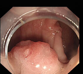
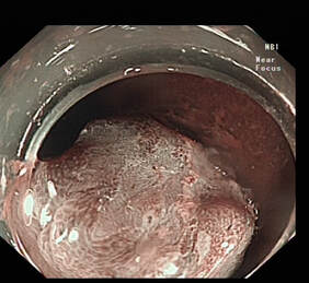
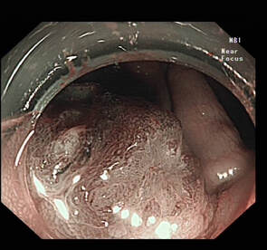
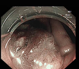
This small 15 mm sigmoid polyp was referred for Endoscopic resection. Patient was fit and well with no comorbidities. What do you think of the surface pattern? Would you offer Endoscopic resection or surgery for this lesion ? What is the predicted histology?
​
The question is how reliably can you trust the surface pattern? And how much importance do you give to this in actual decision making. I classified this as JNET 3 verging on 2B , but as the other factors and dynamic assessment and lifting were favourable , went ahead with ESD for this lesion. What was the eventual histology?
  1. TVA with LGD
  2. TVA with HGD
  3. Adenocarcinoma with early Sm invasion (<1000 )
  4. Adenocarcinoma with deep sm invasion (>1000 )
Picture
Picture
Picture
Picture
The lesion was resected Enbloc with ESD - suboptimal lift - specimen size 25 mm x 25 mm. The histology was one of Pt1B Sm invasive adenocarcinoma, moderately differentiated, with depth of invasion at 2.9 mm beyond muscularis mucosa (Deep sm invasion ) with Lymphovascular invasion. Clear deep margin by 0.9 mm , clear lateral margins. He was offered surgery after this due to LVI
Often experts consider other factors such as
  1. Patient factors- wishes /comorbidities/attitudes
  2. Location of the lesion- access for ER and type of surgery 
  3. Expertise available in advanced resection techniques- such as ESD / Full thickness
  4. Dynamic assessment of lesion in relation to peristalsis/movement etc
  5. Morphological pattern- Paris classification, size
  6. Behaviour of polyp during resection- lifting etc
  7. Gut feeling often drives experts to make a decision and sometime “fit” in the other features accordingly, when the other features are borderline
JNET classification was proposed in 2014. There is limited evidence on validity of JNET and these studies are mainly amongst Japanese endoscopists. Although the sensitivities and specificities for the distinction between benign (1+2A) and malignant (2B+3) groups were 90.0% and 90.0% among experts, and 87.6% and 78.3% among non -GI trainee s, the average diagnostic accuracy was 76.6% for the experts and 61.4% for the trainees in this study (1). To confuse matters polyps often have mixed surface patterns and incompletely visualising the surface may significantly influence interpretation.
 
In conclusion , although its fair to use surface pattern assessment to differentiate malignant and non malignant polyps, but a decision on endoscopic resectability should be based on various factors than just based on JNET Classification.
  1. Minoda Y, Ogino H, Chinen T, Ihara E, Haraguchi K, Akiho H, Takizawa N, Aso A, Tomita Y, Esaki M, Komori K, Otsuka Y, Iwasa T, Ogawa Y. Objective validity of the Japan Narrow-Band Imaging Expert Team classification system for the differential diagnosis of colorectal polyps. Dig Endosc. 2019 Sep;31(5):544-551.
                                                                                                                                                                                   Post by Dr Pradeep Mundre
Comments

    Categories

    All
    Barrett's
    Cancer
    Cancer Syndromes
    Colitis
    Colorectum
    Corrosive Ingestion
    Crypt Pattern
    Difficulty: Hard
    Difficulty: Moderate
    Difficulty: Very Hard
    Duodenum
    Eosinophilic Oesophagitis
    EUS
    Foveolar Metaplasia
    Gastric
    Gastroscopy
    GI Bleeding
    Histology
    HPB
    Ileum
    Immunosuppression
    Infection
    Local Recurrence Of Barrett's Ca.
    Lymphoma
    Mixed Polyp
    Mucosal Prolapse Syndrome
    NET
    Non-lifting Sign
    Oesophagus
    Opinion Piece!
    Pharynx
    Polyp
    Polypectomy
    The Basics
    TSA

  • Home
  • Quiz Cases
  • Endoscopy Long Cases
  • Short Teaching Clips
  • Blackboard teaching
  • Podcasts
  • Core Reading
    • Basic concepts core reading
    • Gastroscopy core reading
    • Colonoscopy core reading
    • QA core reading
  • About
    • Our sponsors