|
Monz found this lesion in the terminal ileum. WHAT IS THE MOST LIKELY DIAGNOSIS?
■ Hyperplastic polyp
Nope, those are very rare in TI
■ Adenomatous polyp
Uncommon in TI and there are no adenomatous crypts!
■ Neuroendocrine tumour
Perhaps the most common lesion in TI with those typical little vessels!
■ Small leiomyoma
Could be but those little vessels say otherwise
■ Small GIST
Must exist but I've never seen one!
explanation
At diagnostic colonoscopies, I do try to intubate the terminal ileum. Not to 'practise' but to detect pathology such as this rare lesion at an early stage! These do have a real malignant potential and in fact usually present late with bowel obstruction.
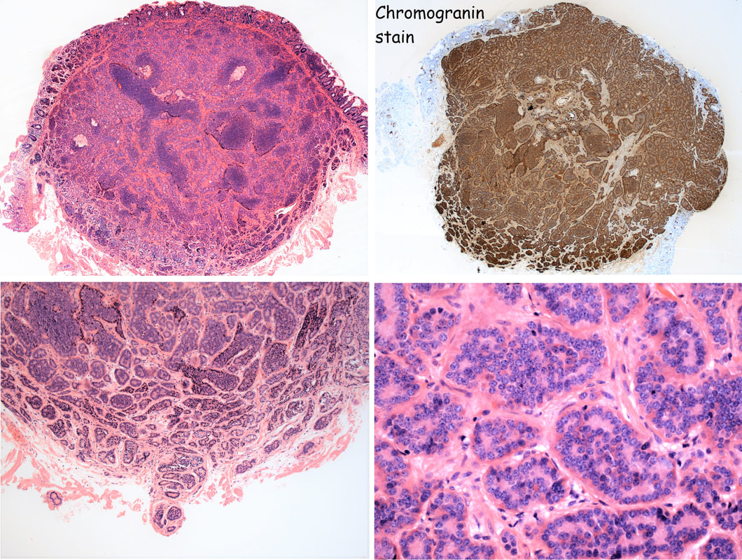
The tell-tale sign are those tiny vessels climbing up around the edges of the polyp and the pale appearance. Of course, this is a terminal ileal NET! The lesion was removed by Monz Ahmed and histology confirmed that it was an NET. Of course, the MOST IMPORTANT issue is the WHO Grade of the lesion. This is because small bowel NET's (excluding the duodenum) are often malignant. The lesion was WHO Grade I (i.e. 1-2% mitotic figures only) which is reassuring but a CT is probably still warranted to look at those nearby lymphnodes. By the way, in the histology below, you can see how close the lesion is on the abutting deep edge. This is the common finding after your endoscopic removal and can be disconcerting. Studies have reported that if you resect lesions using a 'banding device' you can usually get a confirmed (but shallow) clear deep margin. However, neither Monz or I would use a banding device in the terminal ileum. The muscle propria layer is too thin and you are likely to end up with a full thickness resection if you tried. 'Banding resections' are probably best reserved for the oesophagus, stomach or colorectum! |
Categories
All
|

